39 sarcomere diagram labeled
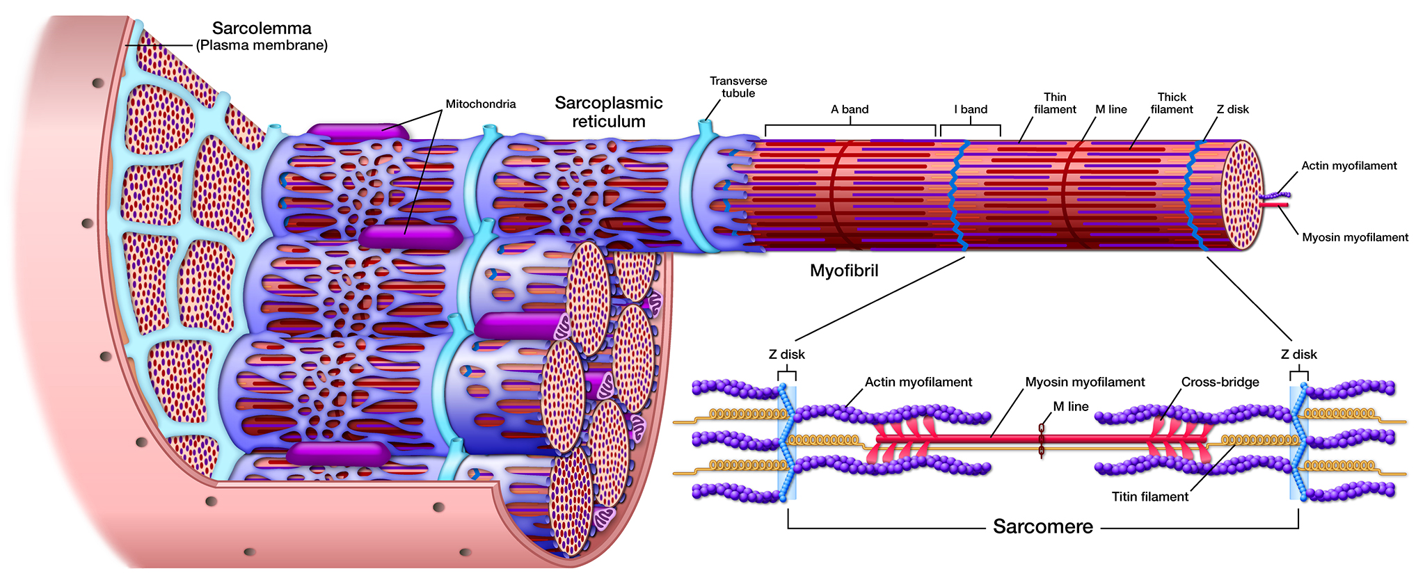
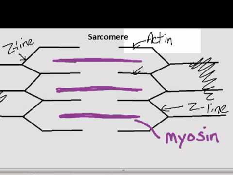
Sarcomere Diagram Labeled - schematron.org Sarcomere Diagram Labeled 16.08.2018 5 Comments Sarcomeres are composed of thick filaments and thin filaments. The thin filaments Look at the diagram above and realize what happens as a muscle contracts. Draw your own diagram of two sarcomeres. The first should be of a relaxed muscle. The second should be of a contracted muscle. 10.2 Skeletal Muscle - Anatomy & Physiology A sarcomere is defined as the region of a myofibril contained between two cytoskeletal structures called Z-discs (also called Z-lines), and the striated appearance of skeletal muscle fibers is due to the arrangement of the thick and thin myofilaments within each sarcomere ( Figure 10.2.2 ).
Sarcomere Thin Filaments Diagram - 16 images [Sarcomere Thin Filaments Diagram] - 16 images - more on the elements of the flesh sarco sarcolemma plasma membrane, muscle anatomy physiology, the thin filaments inward increasing overlap of the filaments and, skeletal muscle tissue,

Sarcomere diagram labeled
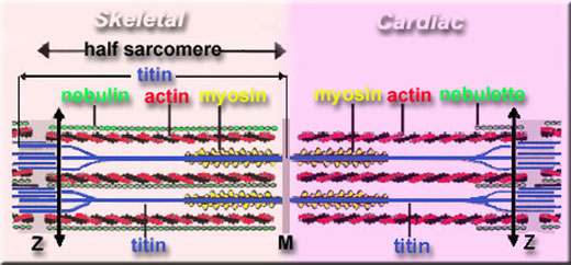
Draw the diagram of a sarcomere of skeletal muscle showing ... - SaralStudy Popular Questions of Class 11 Biology. Q:-Describe briefly the four major groups of Protozoa.Q:-Why are living organisms classified?Q:-State two economically important uses of: (a) Heterotrophic bacteria (b) ArchaebacteriaQ:-What is the difference between direct and indirect development?Q:-Define a taxon. Give some examples of taxa at different hierarchical levels. Anatomy of the cardiac sarcomere. (A) Diagram of the basic organization ... The sarcomere forms the basic contractile unit in the cardiomyocytes of the heart. Thin filaments composed of actin are anchored at the Z line and form transient sliding interactions with thick ... Sarcomere - Definition, Structure, Function and Quiz | Biology Dictionary A sarcomere is the functional unit of striated muscle. This means it is the most basic unit that makes up our skeletal muscle. Skeletal muscle is the muscle type that initiates all of our voluntary movement. Herein lies the sarcomere's main purpose. Sarcomeres are able to initiate large, sweeping movement by contracting in unison.
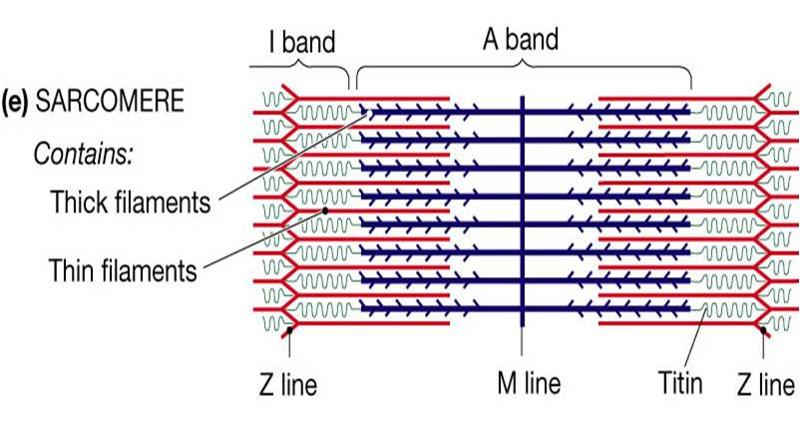
Sarcomere diagram labeled. Solved 12. Label the following diagram of a sarcomere | Chegg.com Label the following diagram of a sarcomere (filaments, bands, lines, etc., ...) ***Which regions shorten during contraction? 13. Describe four actions of ATP in the excitation-contraction coupling in a skeletal muscle fiber. 12-1. 12-2 12-3. 12-4. 14. The neuron and all the muscle fibers it excites are referred to as a 15. Label the Sarcomere Structure Diagram | Quizlet Only $35.99/year Label the Sarcomere Structure STUDY Learn Write Test PLAY Match Created by jack_burton76PLUS Terms in this set (12) z disc mysosin (thick) thin (actin) filament I band A band I band H zone elastic (titin) filaments elastic (titin) filaments thin (actin) filament thick (myosin) filaments myosin heads Sets found in the same folder Draw the diagram of a sarcomere of skeletal muscle class 11 ... - Vedantu Draw the diagram of a sarcomere of skeletal muscle showing different regions. Hint: Sarcomere is the essential unit of striated tissue in the muscles. This means that it is the most important entity that makes up our skeletal muscle. It forms the unit which repeats between two Z lines. By contracting in unison, sarcomeres can initiate broad ... Sarcomere: anatomy, structure and function | Kenhub The structure of the sarcomere is traditionally described with dark and light bands visible under the microscope. This banding pattern in sarcomeres is due mainly to the arrangement of thick and thin myofilaments in each unit. These markings include: A bands (or anisotropic bands) - dark bands that contain whole thick filaments (myosin).
Sarcomere - Wikipedia A sarcomere (Greek σάρξ sarx "flesh", μέρος meros "part") is the smallest functional unit of striated muscle tissue. It is the repeating unit between two Z-lines. Skeletal muscles are composed of tubular muscle cells (called muscle fibers or myofibers) which are formed during embryonic myogenesis. Muscle fibers contain numerous tubular myofibrils. Sarcomere Diagram Labeled - Wiring Diagrams Draw your own diagram of two sarcomeres. The first should be of a relaxed muscle. The second should be of a contracted muscle. Label the Z line, M line. Their observations led to the discovery of sarcomere zones. Sarcomere The figure depicts the structure of a Sarcomere. (Each zone is labeled). They first. Contracted Sarcomere Diagram - Wiring Diagrams The diagram above shows a partially contracted muscle where there is more overlapping of the. Draw your own diagram of two sarcomeres. The first should be of a relaxed muscle. The second should be of a contracted muscle. Label the Z line, M line. (B) A conceptual diagram representing the connectivity of molecules within a sarcomere. DOCX Label the diagram of the skeletal muscle with the following terms ... Label the diagram of the muscle fiber below with the following terms: actin (thin filaments), neuromuscular junction, myofibril, myosin (thick filaments), sarcolemma, sarcomere, sarcoplasmic reticulum, T-tubule, Z-line. 3. Color the muscles of the head and neck in the diagrams below. 4. Color the muscles of the upper back and chest in the ...
Schematic diagram of a muscle sarcomere. The isotropic and anisotropic ... Schematic diagram of a muscle sarcomere. The isotropic and anisotropic bands are labeled as the I-Band and A-Band, respectively. One sarcomere is the length from one Z-Line to the next. Question Video: Identifying the Z Line in the Sarcomere | Nagwa Two successive Z lines mark the boundary of each sarcomere. The A bands, labeled here with the letter Z, are the regions of the sarcomere that do contain the thicker, darker myosin filaments. And so they are sometimes called the dark bands. The outer edges of the A bands are darkest as they contain both actin and myosin filaments overlapping. Sarcomere - Muscle Contraction - SmartDraw Muscle Contraction Skeletal muscle, also called striated muscle tissue, is made up of a series of sarcomeres A sarcomere consists of myosin and actin filaments which overlap upon contraction Myosin bonds with actin to ratchet the tropomyosin down the length of the myosin. Sarcomere Actin filament Troponin Tropomyosin Myosin filament Illustrate sarcomere with a diagram. - Toppr Ask Click here👆to get an answer to your question ️ Illustrate sarcomere with a diagram.
Medical Images | Art & Science Graphics
Labeled Sarcomere Diagram - schematron.org Sarcomeres are composed of thick filaments and thin filaments. The thin filaments Look at the diagram above and realize what happens as a muscle contracts. Each myofibril is made up of contractile sarcomeres AND Drawing labelled diagrams of the structure of a sarcomere. A sarcomere is the basic unit of striated muscle tissue.
Print Anatomy Exam 2 flashcards | Easy Notecards
Sarcomere Labeling Quiz - PurposeGames.com This is an online quiz called Sarcomere Labeling. There is a printable worksheet available for download here so you can take the quiz with pen and paper. Your Skills & Rank. Total Points. 0. ... Label Parts of the Skull - Lateral View 10p Image Quiz. Skeletal Muscle Matching - Front of the Body 14p Image Quiz. Bones of the Skull ...

Skeletal Muscle Tissue
Sarcomere: Structure and Parts, Functions and Histology The main components of the histology of a sarcomere are summarized below: Band A Thick filament zone, composed of myosin proteins. Zone H Central zone of band A, without actin proteins superimposed when the muscle is relaxed. Band I Zone of thin filaments, composed of actin proteins (without myosin). Z disks

Sarcomere Structure and Function - YouTube
Diagram Of Sarcomere The sarcomere is the contractile unit of muscle. This means it is the part of muscle Diagram of the Sarcomere. Source: . A dark stripe called a Z disc marks the ends of one sarcomere and the beginning Look at the diagram above and realize what happens as a muscle contracts.Mass Haul Diagram Explained. Whirlpool Duet Dryer Parts Diagram.
Sarcomere | Cell and Developmental Biology | SUNY Upstate Medical ...
Sarcomere | Definition, Structure, & Sliding Filament Theory The sarcolemma is the thin clear sheath which wrapped the fibers of skeleton muscles. It is a cell membrane which covered striated muscles cells. The sarcolemma is also known as myolemma. The sarcolemma is such as a plasma membrane but specialized in muscle fiber cells. Sarcomere Structure:

neuromuscular junction labeled muscle contraction relaxation notes 3 ...
Labeled Sarcomere Diagram A sarcomere is the basic unit of striated muscle tissue. It is the repeating unit between two Z lines. Skeletal muscles are composed of tubular muscle cells which. Sarcomeres are composed of thick filaments and thin filaments. The thin filaments Look at the diagram above and realize what happens as a muscle contracts.
.jpg)
This figure shows the structure of the muscle fibers. In the top panel ...
Sarcomere (Muscle) Coloring - The Biology Corner The enter muscle fiber is surrounded by the sarcolemma (D), color this membrane brown. If expanded, the light and dark bands are shown as individual thick and thin filaments. Color the thick filaments (not labeled) red and the thin filaments blue. The Z line is the boundary between sarcomeres, named after its shape. Color the Z-line orange. 1.
Parts of the Sarcomere - YouTube
Question Video: Recalling the Part of the Sarcomere that ... - Nagwa The diagram shows a labeled structure of a sarcomere. Which part contains actin and myosin? To answer this question, let's start by addressing what a sarcomere is before we look at its structure in more detail. A sarcomere is the functional unit of organelles found exclusively in muscle cells called myofibrils.
Post a Comment for "39 sarcomere diagram labeled"