42 drag the labels to the correct locations on these images of human chromosomes.
Cell Cycles: Interphase, Mitosis, Cytokinesis - SchoolWorkHelper Cell Cycle: nuclear division, cytokinesis Parental cell: genetic copies of parental cell 3 process: checks/ regulators for each step to ensure timely progression, replication process to synthesis DNA into two copies, interwoven "cables" and "motors" of mitotic cytoskeletons. Chromosomes: nuclear units of genetic information; DNA molecules combined with proteins. In eukaryotes, heredity ... Drag the labels to the correct locations on these images of human ... Drag the labels to the correct locations on these images of human chromosomes - Brainly.com. bugz337owsm7z. 09/24/2017. Biology.
Cell division: mitosis and meiosis | Biological Principles These mitotic chromosomes each consist of a pair of sister chromatids joined at their centromeres. The images of the homologous chromosome pairs (e.g., 2 copies of chromosome 1) have been lined up next to each other. Image from Bolzer et al., (2005) Three-Dimensional Maps of All Chromosomes in Human Male Fibroblast Nuclei and Prometaphase Rosettes.

Drag the labels to the correct locations on these images of human chromosomes.
Stages Of Meiosis Diagram Labeled - Wiring Diagrams Drawing diagrams to show the stages of meiosis resulting in the formation of four haploid cells. Meiosis consists of two divisions, both of which follow the same stages as mitosis (prophase, metaphase, anaphase, telophase) P-I: Chromosomes condense, nuclear membrane dissolves.Mitosis: Labeled Diagram Mitosis is a process of cell division which ... Chromosomes Fact Sheet - Genome.gov Chromosomes are thread-like structures located inside the nucleus of animal and plant cells. Each chromosome is made of protein and a single molecule of deoxyribonucleic acid (DNA). Passed from parents to offspring, DNA contains the specific instructions that make each type of living creature unique. Phases of mitosis | Mitosis | Biology (article) | Khan Academy Mitosis consists of four basic phases: prophase, metaphase, anaphase, and telophase. Some textbooks list five, breaking prophase into an early phase (called prophase) and a late phase (called prometaphase). These phases occur in strict sequential order, and cytokinesis - the process of dividing the cell contents to make two new cells - starts ...
Drag the labels to the correct locations on these images of human chromosomes.. BYJUS Chapter 8 Mastering Flashcards - Quizlet Explanation: Each duplicated chromosome consists of two identical sister chromatids. Drag the labels onto the diagram to identify the stages of the cell cycle. Most of the cell's life is spent in interphase, when growth occurs. Cells are about to divide and replicate their DNA The Cell Cycle | Biology I | | Course Hero Spindle microtubules that do not engage the chromosomes are called polar microtubules. These microtubules overlap each other midway between the two poles and contribute to cell elongation. Astral microtubules are located near the poles, aid in spindle orientation, and are required for the regulation of mitosis. Chapter 8 Homework Flashcards - Quizlet Drag the labels to the correct locations on these images of human chromosomes. Drag the labels onto the diagram to identify the stages of the life cycle. (Drag only blue labels onto blue targets and pink labels onto pink targets.) The function (s) of meiosis is/are _____. reproduction (production of gametes)
Solved Drag the labels to the correct locations on these - Chegg Drag the labels to the correct locations on these images of human chromosomes. Show transcribed image text Expert Answer 95% (21 ratings) a. homologous chromos … View the full answer Transcribed image text: centromere d autosomes (с . sister chromatids karyotype e sex chromosomes homologous chromosomes Photograph by CNRISPLPhoto Researchers 6.2 The Cell Cycle - Concepts of Biology - 1st Canadian Edition Cells on the path to cell division proceed through a series of precisely timed and carefully regulated stages of growth, DNA replication, and division that produce two genetically identical cells. The cell cycle has two major phases: interphase and the mitotic phase ( Figure 6.3 ). During interphase, the cell grows and DNA is replicated. PDF ORGANELLE LOCATION DESCRIPTION FUNCTION - Greeley Schools These are small-sized sac like structures. They are of different types lysosomes, peroxisomes. *These help in storage and release of substances as required by the cell. For example lysosomes help in cell digestion when cell dies. Vacuoles function is to store water. lysosome plant - uncommon animal - common A Labeled Diagram of the Plant Cell and Functions of its Organelles The nucleus is known to be the 'control room' of the cell. It regulates various cell functions by controlling the protein synthesis of the plant cell. The nucleus contains DNA within the chromosomes. It is a membrane-bound structure that contains the cells hereditary information. Function: Controls expression and transcription of the gene ...
9.2 DNA Replication - Concepts of Biology - 1st Canadian Edition When a cell divides, it is important that each daughter cell receives an identical copy of the DNA. This is accomplished by the process of DNA replication. The replication of DNA occurs during the synthesis phase, or S phase, of the cell cycle, before the cell enters mitosis or meiosis. The elucidation of the structure of the double helix ... A Labeled Diagram of the Animal Cell and its Organelles In humans, X and Y are sex chromosomes. Females have two X chromosomes and males have one X and one Y chromosome. Autosomes are all the other chromosomes in the organism. Of the 46 chromosomes in humans, 44 are autosomes and the remaining two are the sex chromosomes. Can You Correctly Label These Images Of Chromosomes Drag the labels to the correct locations on these images of human chromosomes. Of the 46 chromosomes in humans 44 are autosomes and the remaining two are the sex chromosomes. PROPHASE I Homologous chromosomes pair up and form tetrad 2. Drag the labels onto the diagram to identify the stages of the life cycle. Lab Manual Exercise #2 - Palomar College Homologous Chromosomes: Paternal and Maternal Maternal (pink) and paternal (blue) sets of chromosome doublets. Two pairs of homologous chromosome doublets. Maternal and paternal sets of single chromosomes. Single chromosomes and doubled chromosomes (chromosome doublets). Beginning with prophase, the chromosomes appear as doublets.

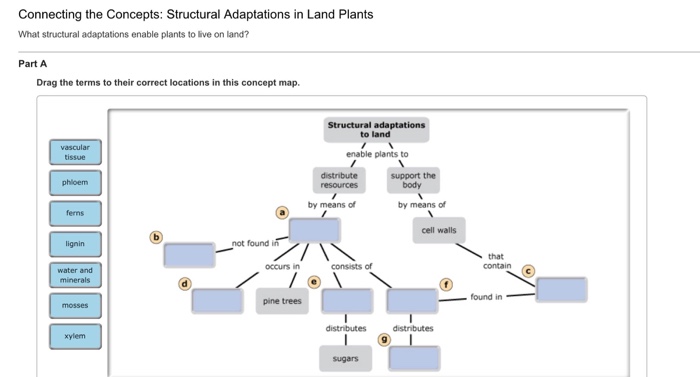
Solved: Drag The Terms To Their Correct Locations In This ... | Chegg.com
Mastering Biology 5 Flashcards - Quizlet All the offspring of a cross between a black-eyed cat and an orange-eyed cat have black eyes. This means that the allele for black eyes is __________ the allele for orange eyes. Mendel formulated his principles of inheritance based on _____. observations on the outcomes of breeding experiments.
Biology Archive | December 19, 2016 | Chegg.com
Mastering Biology - Chapters 10 and 11 Assignment - Quizlet Drag the labels to the correct locations on these images of human chromosomes. a. homologous chromosomes b. centromere c. sister chromatids d. autosomes e. sex chromosomes f. karyotype. Drag the labels onto the diagram to identify the stages of the life cycle. Not all labels will be used.
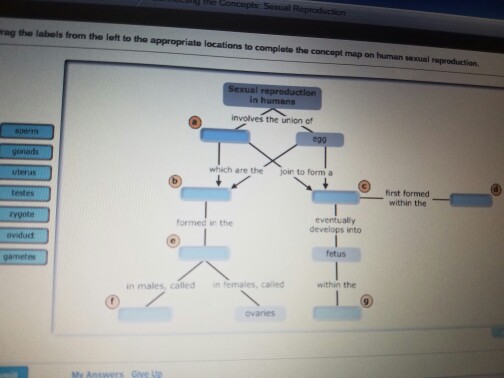
Drag The Terms To Their Correct Locations In This Concept Map About ...
ch 8 mastering biology Flashcards - Quizlet Drag the labels onto the diagram to identify the stages of the cell cycle. 1. most of the cells life is spent in interphase 2. in phosphase microtubules form the mitototic spindle 3. at metaphase, the mitotic spindle is fully formed 4. in anaphase, sister chromatides separate 5. in telophase chromosomes become less condenced

Solved: Drag The Labels From The Left To Their Correct Loc... | Chegg.com
Solved Learning through Art: Chromosomes Part A Drag the - Chegg Learning through Art: Chromosomes Part A Drag the labels to the correct locations on these images of human chromosomes. Reset Help Sister chromatids chromosomes autosomes centromere homologous chromosomes karyotype Photograph by CRISP Photosmarchers Submit Request Answer

Solved: Can You Correctly Place The Labels In This Diagram... | Chegg.com
Karyotyping Activity - University of Arizona During mitosis, the 23 pairs of human chromosomes condense and are visible with a light microscope. A karyotype analysis usually involves blocking cells in mitosis and staining the condensed chromosomes with Giemsa dye. The dye stains regions of chromosomes that are rich in the base pairs Adenine (A) and Thymine (T) producing a dark band.

Biology Archive | April 18, 2016 | Chegg.com
Phases of mitosis | Mitosis | Biology (article) | Khan Academy Mitosis consists of four basic phases: prophase, metaphase, anaphase, and telophase. Some textbooks list five, breaking prophase into an early phase (called prophase) and a late phase (called prometaphase). These phases occur in strict sequential order, and cytokinesis - the process of dividing the cell contents to make two new cells - starts ...

34 Can You Correctly Label The Structures In This Diagram That ...
Chromosomes Fact Sheet - Genome.gov Chromosomes are thread-like structures located inside the nucleus of animal and plant cells. Each chromosome is made of protein and a single molecule of deoxyribonucleic acid (DNA). Passed from parents to offspring, DNA contains the specific instructions that make each type of living creature unique.
32 Can You Label These Chromosomes With The Correct Genetic Terms ...
Stages Of Meiosis Diagram Labeled - Wiring Diagrams Drawing diagrams to show the stages of meiosis resulting in the formation of four haploid cells. Meiosis consists of two divisions, both of which follow the same stages as mitosis (prophase, metaphase, anaphase, telophase) P-I: Chromosomes condense, nuclear membrane dissolves.Mitosis: Labeled Diagram Mitosis is a process of cell division which ...
Post a Comment for "42 drag the labels to the correct locations on these images of human chromosomes."