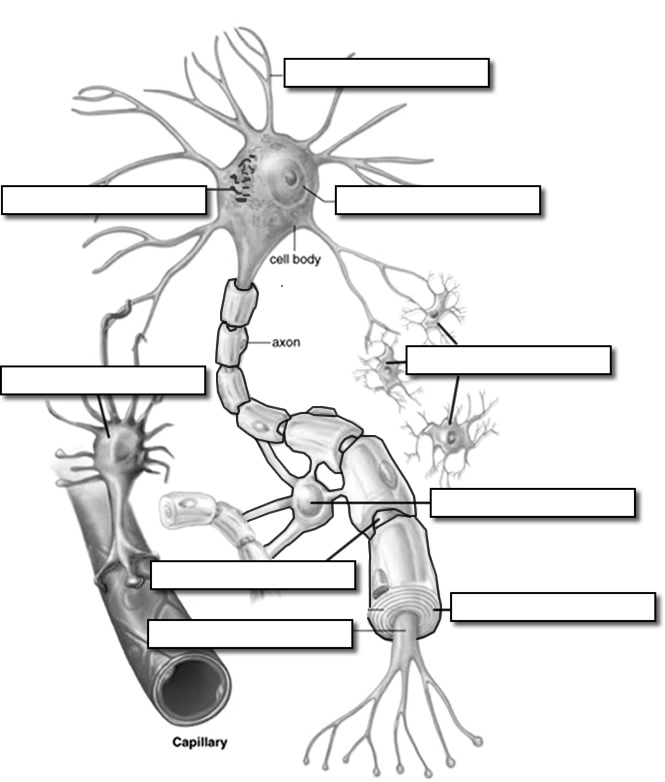
45 unlabeled neuron diagram
Artificial networks learn to smell like the brain Unlabeled diagram of the olfactory system showing the anatomy of smell. ... who reported their findings Oct. 6 in the journal Neuron, say their artificial network will help researchers learn more ... Diagram of Human Heart and Blood Circulation in It Four Chambers of the Heart and Blood Circulation. The shape of the human heart is like an upside-down pear, weighing between 7-15 ounces, and is little larger than the size of the fist. It is located between the lungs, in the middle of the chest, behind and slightly to the left of the breast bone. The heart, one of the most significant organs ...
Animal Cell- Definition, Structure, Parts, Functions, Labeled Diagram An animal cell is a eukaryotic cell that lacks a cell wall, and it is enclosed by the plasma membrane. The cell organelles are enclosed by the plasma membrane including the cell nucleus. Unlike the animal cell lacking the cell wall, plant cells have a cell wall. Animals are a large group of diverse living organisms that make up three-quarters ...
Unlabeled neuron diagram
Anatomy of the eye: Quizzes and diagrams | Kenhub Take a look at the diagram of the eyeball above. Here you can see all of the main structures in this area. Spend some time reviewing the name and location of each one, then try to label the eye yourself - without peeking! - using the eye diagram (blank) below. Unlabeled diagram of the eye. Click below to download our free unlabeled diagram of ... Nervous system: Structure, function and diagram | Kenhub Neurons, or nerve cell, are the main structural and functional units of the nervous system.Every neuron consists of a body (soma) and a number of processes (neurites). The nerve cell body contains the cellular organelles and is where neural impulses (action potentials) are generated.The processes stem from the body, they connect neurons with each other and with other body cells, enabling the ... Animal Cell And Label - Blank Animal Cell Diagram To Label Ythoreccio ... Terms in this set (12) centriole. Organelle that helps with cell division. Some of the worksheets for this concept are lesson life science plant animal cell functions, cell diagram to label activity, name animal cell vs plant cell, plant and animal cell labeling diagram, plant labeling work, lesson 5 plant and animal cells, plant and animal.
Unlabeled neuron diagram. Toward a More Accurate 3D Atlas of C. elegans Neurons The. 30. goal of this paper is to further improve the scienti fi c utility of C. elegans by enabling more accur ate. 31. and automated identi fi cation of its neurons, that are either unlabeled ... SANTIA: a Matlab-based open-source toolbox for artifact detection and ... The diagrams of the main components of these architectures are depicted in Fig. 3. The LSTM architecture is a type of recurrent network spanning adjacent time steps in a manner that at every point the neurons take the current data input as well as the values of the hidden neurons that collect the information of the previous time steps. Anterograde or retrograde transsynaptic labeling of CNS neurons with ... Upstream neurons were optically activated with pulses of blue light. (G) Diagram of recording configuration ... synaptic connectivity was observed in 5/8 pairs of infected neurons vs. 0/10 pairs where one neuron was unlabeled. It is likely that greater than 5/8 of the pairs were connected, as there are multiple reasons why currents may not have ... A Complete Guide To Artificial Neural Network In Machine Learning Basic Models Of ANN. Neural Network Architecture. #1) Single-Layer Feed-Forward Network. #2) Multi-Layer Feed-Forward Network. #3) Single Node With Its Own Feedback. #4) Single Layer Recurrent Network. #5) Multi-Layer Recurrent Network. Example Of Artificial Neuron Network. Comparison Between Machine Learning And ANN.
Learn all muscles with quizzes and labeled diagrams | Kenhub Human body muscle diagrams. Muscle diagrams are a great way to get an overview of all of the muscles within a body region. Studying these is an ideal first step before moving onto the more advanced practices of muscle labeling and quizzes. If you're looking for a speedy way to learn muscle anatomy, look no further than our anatomy crash courses . Artificial neural network - Wikipedia An artificial neural network is an interconnected group of nodes, inspired by a simplification of neurons in a brain. Here, each circular node represents an artificial neuron and an arrow represents a connection from the output of one artificial neuron to the input of another. Artificial neural networks ( ANNs ), usually simply called neural ... Principles of connectivity among morphologically defined cell types in ... Importantly, each type of neuron has its own characteristic input-output connectivity profile, connecting with other constituent neuronal types with varying degrees of specificity in postsynaptic targets, laminar location, and synaptic characteristics. ... All unlabeled neurons recorded from L23 (n = 120) and L5 ... Wiring diagram of V1 ... Human Body Diagram Worksheet Body diagram worksheet a muscle diagram unlabeled skull. Get it has a friend made by visitors, called human heart worksheets with the metatarsals, body human diagram worksheet images are illustrated science with a normal force or. ... Neuron diagram blank human body anatomy human body anatomy. ICU, intubation or mechanical ventilation, or death.
Nervous System: TEAS - || RegisteredNursing.org The nervous system is comprised of an intricate system of millions of nervous system cells which are called neurons and glia cells. The neuron is the primary type of cell in the nervous system. They are, in fact, often referred to as a nerve cell. Glial cells, on the other hand, are a type of nervous system cell, other than a neuron, that forms ... Neuron secrete exosomes containing miR-9-5p to promote polarization of ... e Representative NIRF images of brains from the DiR-labeled group exosomes-treated and unlabeled exosomes-treated group. f ... morphology of primary neuron. Scale bar = 50 μm. (Right) The PCR results showed that primary neuron like PC12 cells were able to secrete exosome containing miR-9-5p. ... c Venn diagram showed that miR-9-5p was a miRNA ... Structure of Neurons: What Is a Neuron? Types, Structure, Parts Structure of Neuron. Each neuron has a cell body, which is the central area of the neuron. It contains the nucleus and other structures common to all cells in the body, such as mitochondria. Neurons have highly branched fibres that reach out from the neuron are called dendritic trees. Each branch is called a dendrite. Learn anatomy of the scapula with quizzes and diagrams - Kenhub Learn the anatomy of the scapula with quizzes and labeled diagrams. The scapula (AKA: the shoulder blade) is a flat, triangular shaped bone connecting the upper limb with the trunk. Along with the clavicle and manubrium of the sternum, it makes up one of three parts of the pectoral (shoulder) girdle. In this article, we'll be helping you to ...
Neuroscience for Kids - Fill In #1
Artificial networks learn to smell • tectales • tagging medical technology Using machine learning, a computer model can teach itself to smell in just a few minutes. When it does, researchers have found, it builds a neural network that closely mimics the olfactory circuits that animal brains use to process odors. Unlabeled diagram of the olfactory system showing the anatomy of smell. Source: Andrewmeyerson/Wikimedia ...
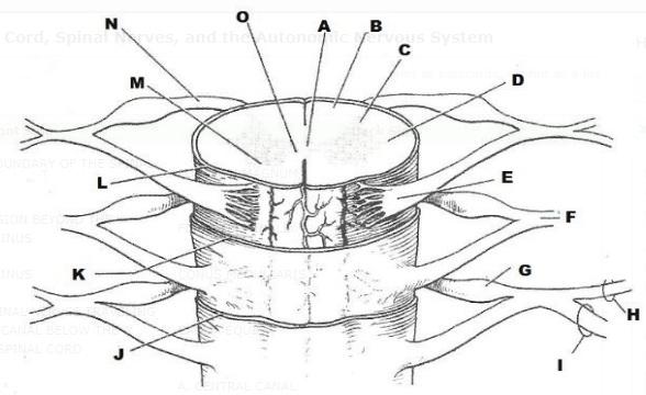
Exercise 21: Spinal Cord, Spinal Nerves, and the Autonomic Nervous ...
Reflex Action: Know the definition, Types, Examples, Diagram - Embibe Fig: Reflex Arc. The reflex arc is the neural pathway that begins with a sensory neuron at a receptor (e.g., a pain receptor in the fingertip) and ends with a motor neuron at an effector (e.g., a skeletal muscle), and as mentioned above, it has following parts: 1. A receptor, which receives stimulus from the surroundings. 2. Afferent or sensory nerve, which takes a sensory impulse to the ...
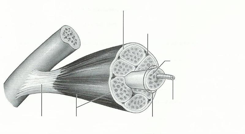
Exercise 14: Microscopic Anatomy and Organization of Skeletal Muscle ...
Plant Cell- Definition, Structure, Parts, Functions, Labeled Diagram Functions of the plant cell (plasma) membrane. In-plant cells the cell membrane separated the cytoplasm from the cell wall. It has a selective permeability hence it regulates the contents that move in and out of the cell. It also protects the cell from external damage and provides support and stability to the cell.

Unlabelled clipart - Clipground
On Optical Detection of Densely Labeled Synapses in Neuropil and ... One can threshold this diagram with certain thresholds, T 1 for the pre-synaptic marker and T 2 for the post-synaptic marker (dashed lines), in order to separate the proximal (red) from distant (blue) voxels, and thus detect presence of a synapse. C) Using correlations in the fluorescence from the pre- and post-synaptic markers, synapses may be ...
muscle, circulatory, + nervous system Flashcards | Easy Notecards
Multipolar Nerve Cell | Function, Structure, Features & Summary Benefits. When considering the structural features of a nerve cell, it is common to speak of a multipolar type of neuron. It possesses a nerve cell body, the perikaryon, from which, as mentioned above, a number of dendritic extensions and one axonal extension originate. As a result, this is the key feature of these nerve cells - a single axon ...
Neuron Label
The Facial Nerve (CN VII) | Cranial Nerves - Geeky Medics The facial nucleus. The facial motor nucleus is a round aggregation of motor neuron cell bodies found in the pontomedullary junction.. These are paired on the left and right side of the brainstem and are neatly divided horizontally in half.. The superior half of the nucleus representing the superior half of the face and the inferior half of the nucleus representing the inferior half of the face.

Brain Labeling Worksheet | Biology worksheet, Cell diagram, Human ...
The Cells and Logic for Mammalian Sour Taste detection - PMC Upper diagram illustrates the strategy used to target DTA or GFP to selective populations of TRCs. ... -positve cells) or unlabeled (control) cells were examined for pH responses. (b) Responses of a sample GFP-labeled or unlabeled neuron to test solutions under a perfusion regime consisting of pH 7.4, pH 6.9, pH 7.4 and pH 6.5; shown are AP ...

Neuron Labeling Quiz
Neuron secrete exosomes containing miR-9-5p to promote polarization of ... Background Neuroinflammation is an important component mechanism in the development of depression. Exosomal transfer of MDD-associated microRNAs (miRNAs) from neurons to microglia might exacerbate neuronal cell inflammatory injury. Results By sequence identification, we found significantly higher miR-9-5p expression levels in serum exosomes from MDD patients than healthy control (HC) subjects ...
Post a Comment for "45 unlabeled neuron diagram"