40 label the structures of a skeletal muscle
Anatomy quizzes - PurposeGames Label the Long Bone by deanne1480 147,367 plays 10p Image Quiz. ... Internal Structures by orkide1 120,839 plays 24p ... Skeletal muscle fiber model by cwalsh2 36,150 ... Muscle Structure Labeling Quiz - PurposeGames.com This online quiz is called Muscle Structure Labeling. This online quiz is called Muscle Structure Labeling. English en. Login. Login Register Free Help; Start; Explore. Games; Playlists; Tournaments; Tags; The Wall; Badges; ... Skeletal Muscle Matching - Back of the Body 10p Image Quiz. Steps of Muscle Contraction 13p Matching Game. Vertebral ...
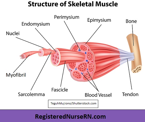
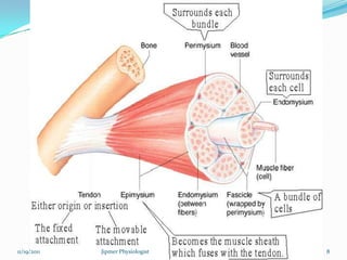
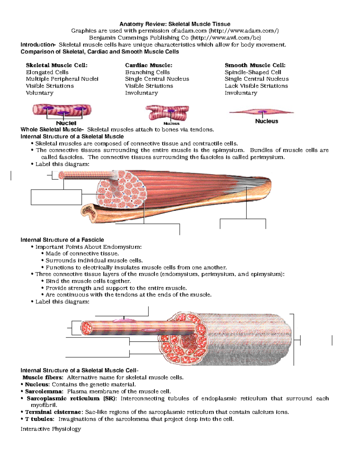
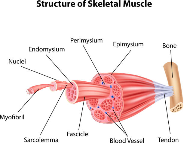
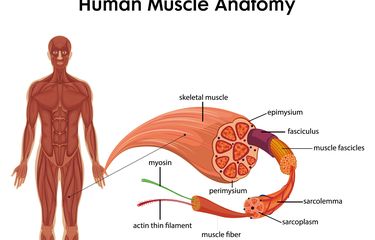
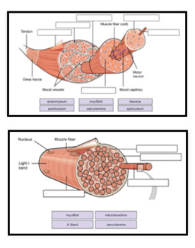
Skeletal Muscle Tissue Anatomy and Structure - Registered Nurse RN Skeletal Muscle Structure Each skeletal muscle is considered an organ, and it's made up of connective tissue layers, muscle fibers, blood vessels, and nerves. Skeletal muscles attach to the bones through tendons or through a direct attachment. As you look at this muscle diagram, you'll notice an outer layer of connective tissue called epimysium.

Label the structures of a skeletal muscle
Naming Skeletal Muscles | Anatomy and Physiology I | | Course Hero The skeletal muscle's anatomical location or its relationship to a particular bone often determines its name. For example, the frontalis muscle is located on top of the frontal bone of the skull. Similarly, the shapes of some muscles are very distinctive and the names, such as orbicularis, reflect the shape. Structure of Skeletal Muscle | SEER Training Each organ or muscle consists of skeletal muscle tissue, connective tissue, nerve tissue, and blood or vascular tissue. Skeletal muscles vary considerably in size, shape, and arrangement of fibers. They range from extremely tiny strands such as the stapedium muscle of the middle ear to large masses such as the muscles of the thigh. Drag each label to the correct location on the diagram. - Brainly.com 1. Muscle: soft, contractile tissue important to produce force and motion in animals. 2. Fascicle: multiple bundles of skeletal muscle fibres which is surrounded by a type of connective tissue called perimysium. 3. Muscle fibres: bundles of cylindrical organelles myofibrils formed by the fusion of myoblasts via myogenesis process. 4.
Label the structures of a skeletal muscle. welcome to Ms. stephens' anatomy and Physiology and … Unit 5: Muscular System Student Learning Goals: I can identify smooth, skeletal, and cardiac muscle tissue under a microscope and state the function of each.; I can identify the component parts of a muscle: fascicle, myofibril, fiber, nucleus of cell, body of muscle.; I can identify the major muscles of the human body.; I can analyze experimental data using the Moving Arm … 108 Muscle Quizzes Online, Trivia, Questions ... - ProProfs Mar 22, 2022 · Here is the amazing physiology of muscle contraction MCQs with answers. Take this quiz if you think you know enough about muscle contraction and physiology. Muscle contraction is the tightening, shortening, or lengthening of... Skeletal Muscle Fiber Structure and Function - Open Textbooks for Hong Kong The striated appearance of skeletal muscle tissue is a result of repeating bands of the proteins actin and myosin that occur along the length of myofibrils. Myofibrils are composed of smaller structures called myofilaments. There are two main types of myofilaments: thick filaments and thin filaments. muscle labeling anatomy Anatomy and Physiology - Wiki - Scioly.org. 9 Images about Anatomy and Physiology - Wiki - Scioly.org : Anatomy of human forearm muscles with labels — Stock Photo | #174714884, Muscular System Review Activity for Muscles | Muscular system and also Connective tissue wrappings of skeletal muscle.
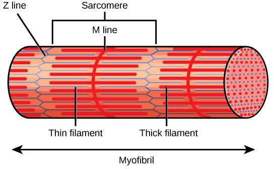
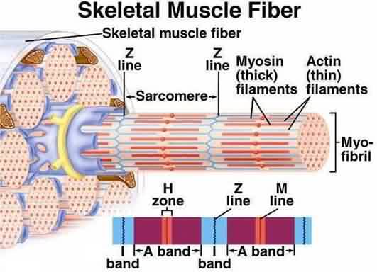
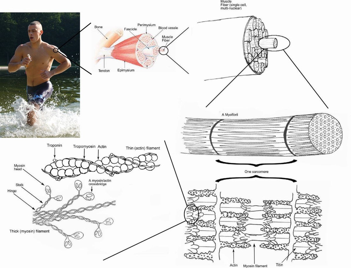
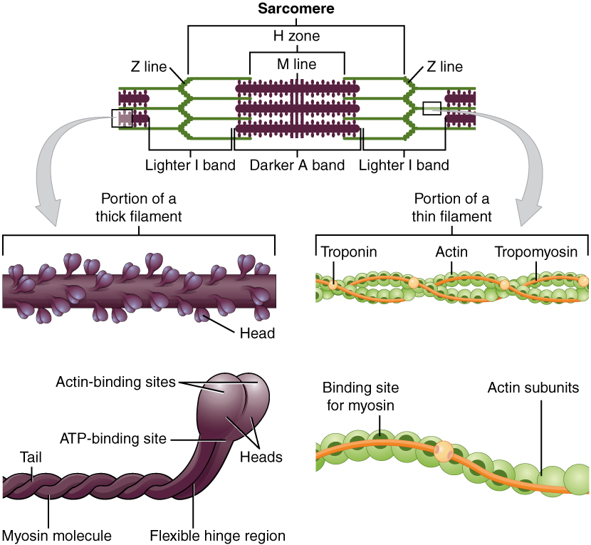
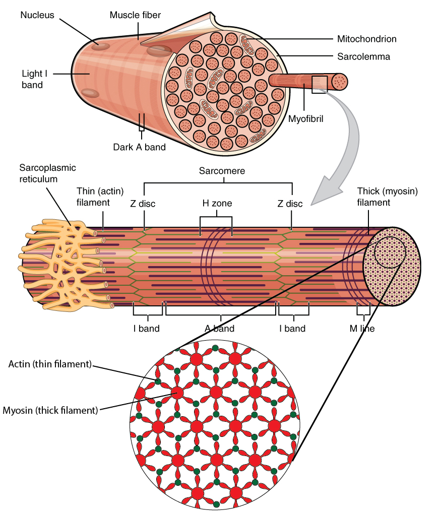
skeletal muscle anatomy worksheet Skeletal Muscle, Diagram - Stock Image - C043/5448 - Science Photo Library . skeletal. 14 best images of muscle labeling worksheets answers and blanks. Diagram muscle blank label human body physiology diagrams anatomy system muscles muscular coloring worksheets drawing quiz worksheet chart major google. Muscle skeletal ... Structure of Skeletal Muscle - CliffsNotes Within a myofibril, actin and myosin filaments are parallel and arranged side by side. The overlapping filaments produce a repeating pattern that gives skeletal muscle its striated appearance. Each repeating unit of the pattern, called a sarcomere, is separated by a border, or Z disc (Z line), to which the actin filaments are attached. Skeletal Muscle - Anatomy & Physiology - University of Hawaiʻi These tissues include the skeletal muscle fibers, blood vessels, nerve fibers, and connective tissue. Each skeletal muscle has three layers of connective tissue (called "mysia") that enclose it and provide structure to the muscle as a whole, and also compartmentalize the muscle fibers within the muscle ( [link] ). DOCX Label the diagram of the skeletal muscle with the following terms ... 2. Label the diagram of the muscle fiber below with the following terms: actin (thin filaments), neuromuscular junction, myofibril, myosin (thick filaments), sarcolemma, sarcomere, sarcoplasmic reticulum, T-tubule, Z-line
Striated muscle: Structure, location, function | Kenhub Skeletal musculature Structure of the skeletal muscle. Muscle fibers and connective tissue layers make up the skeletal muscle.A skeletal muscle fiber is around 20-100 µm thick and up to 20 cm long.Embryologically. it develops by the chain-like fusion of myoblasts. About 200-250 muscle fibers are surrounded by endomysium forming the functional unit of the muscle, the primary bundle. Skeletal muscle tissue: Histology | Kenhub Skeletal muscle is an excitable, contractile tissue responsible for maintaining posture and moving the orbits, together with the appendicular and axial skeletons. It attaches to bones and the orbits through tendons. Excitable tissue responds to stimuli through electrical signals. Contractile tissue is able to generate tension of force. Anatomy quizzes - PurposeGames Label the Skeleton by Mr. Shumaker 893,752 plays 25p Image Quiz. ... Anatomy of the Human Heart - Internal Structures by orkide1 120,839 plays 24p Image Quiz. ... Skeletal muscle fiber model by cwalsh2 36,150 plays 15p Image Quiz. Your Skills & Rank. Total Points. 0. Get started! Today's Rank--0. anatomy of the skeletal muscle Skull - Inferior View - Human Body Help. 11 Pics about Skull - Inferior View - Human Body Help : Simple Muscle Anatomy Simple Muscle Diagram For Kids Label The Major, Skeletal Muscle Anatomy and also Skeletal Muscle Anatomy. Skull - Inferior View - Human Body Help . inferior humanbodyhelp
Skeletal Muscle | Anatomy and Physiology I
Tissue Engineering for Clean Meat Production - Frontiers Increasing public awareness of foodborne illnesses, factory farming, and the ecological footprint of the meat industry, has generated the need for animal-free meat alternatives. In the last decade, scientists have begun to leverage the knowledge and tools accumulated in the fields of stem cells and tissue engineering toward the development of cell-based meat (i.e., clean meat).
SISTEM OTOT MANUSIA
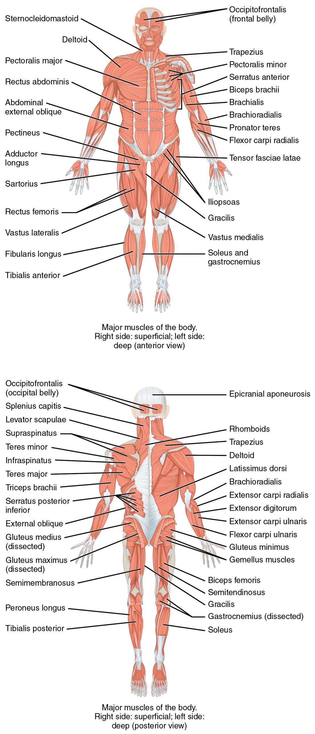
Naming Skeletal Muscles - Anatomy and Physiology The skeletal muscle's anatomical location or its relationship to a particular bone often determines its name. For example, the frontalis muscle is located on top of the frontal bone of the skull. Similarly, the shapes of some muscles are very distinctive and the names, such as orbicularis, reflect the shape.
A skeletal muscle is composed of a variety of tissues - ppt ...
The excitation–contraction coupling mechanism in skeletal muscle Jan 24, 2014 · The most suitable dyes to study ECC in skeletal muscle seem to be the low affinity Ca 2+ dyes, Mag-Fura-2 and Mag-Fluo-4 (Fig. 1), since they are well known and can reliably track fast, large and brief Ca 2+ transients such as those found in skeletal muscle (Hollingworth et al. 2009; Baylor and Hollingworth 2011; Calderón et al. 2010, 2013).

Skeletal Muscle Structure, Illustration Photograph by Spencer ...
Anatomy, Skeletal Muscle - StatPearls - NCBI Bookshelf Each skeletal muscle consists of thousands of muscle fibers wrapped together by connective tissue sheaths. The individual bundles of muscle fibers in a skeletal muscle are known as fasciculi. The outermost connective tissue sheath surrounding the entire muscle is known as epimysium.

Ch 10 lab map Flashcards | Quizlet
Week 6: Muscle Physiology Flashcards & Practice Test - Quizlet Action potential propagation in a skeletal muscle fiber ceases when acetylcholine is removed from the synaptic cleft. Which of the following mechanisms ensures a rapid and efficient removal of acetylcholine?-Acetylcholine is transported back into the axon terminal by a reuptake mechanism.-Acetylcholine is degraded by acetylcholinesterase.

Bellringer 12/9 Label #4 and #5. Building Skeletal Muscle… We ...
welcome to Ms. stephens' anatomy and Physiology and ... Unit 5: Muscular System Student Learning Goals: I can identify smooth, skeletal, and cardiac muscle tissue under a microscope and state the function of each.; I can identify the component parts of a muscle: fascicle, myofibril, fiber, nucleus of cell, body of muscle.
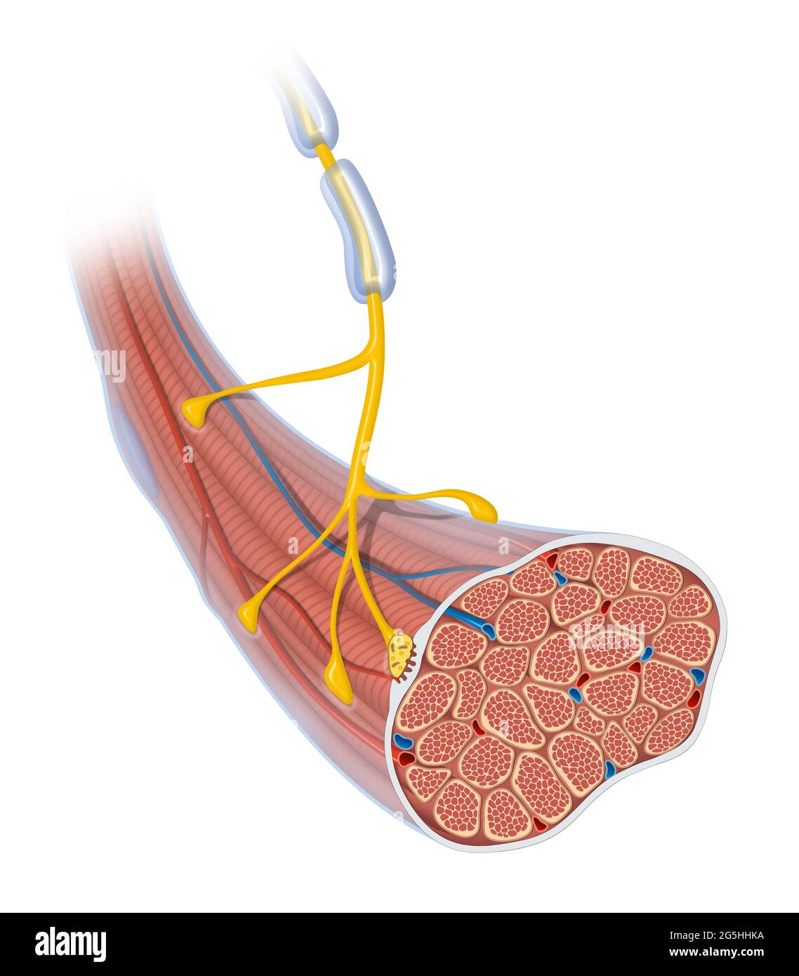
Neuromuscular junction. Skeletal muscle and motor neuron ...
Skeletal Muscle: What Is It, Function, Location & Anatomy The majority of the muscles in your body are skeletal muscles. They make up between 30 to 40% of your total body mass. Tendons (tough bands of connective tissue) attach skeletal muscle tissue to bones throughout your body. Your shoulder muscles, hamstring muscles and abdominal muscles are all examples of skeletal muscles.

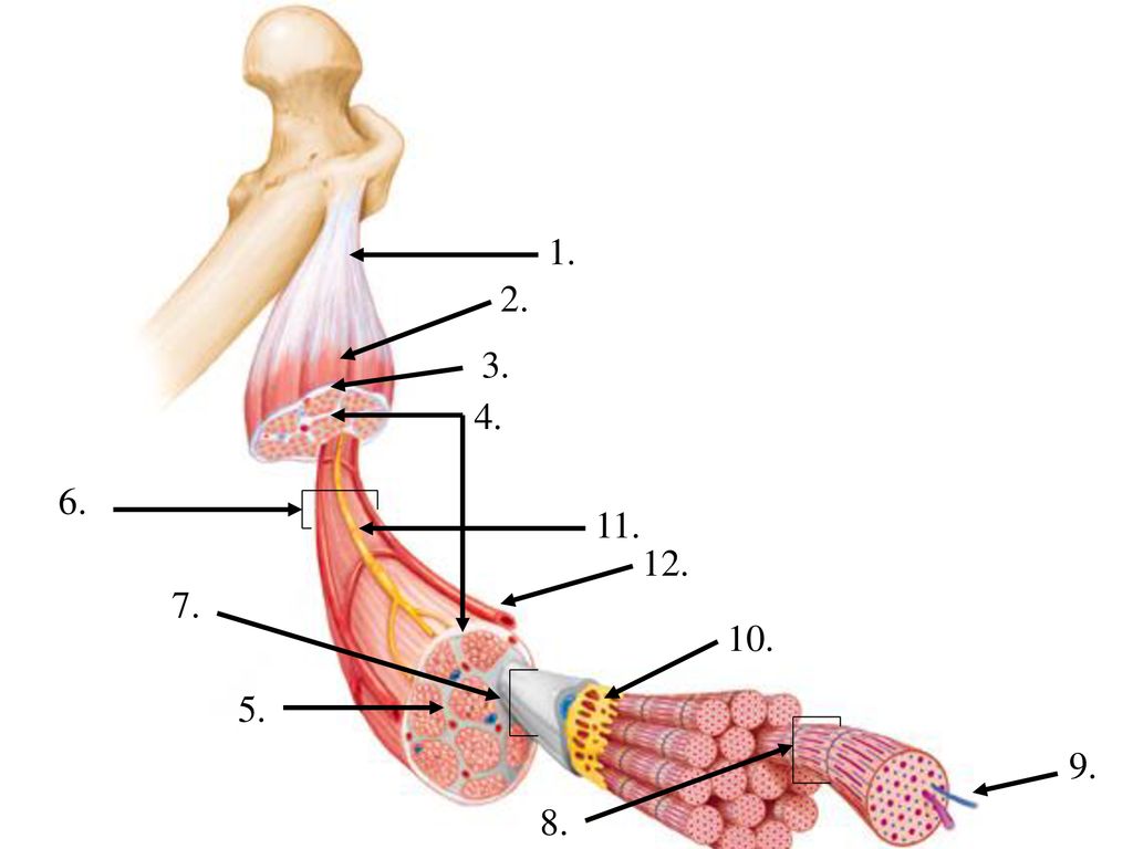
Solved Muscle Cell Label the structures of a skeletal muscle ...
Muscle-derived fibro-adipogenic progenitor cells for ... - Nature 24-01-2022 · Alongside mature multinuclear muscle fibres, skeletal muscle contains a large number of different cell types 47,48. Whilst purifying satellite cells by FACS 34 , we had previously observed a large ...
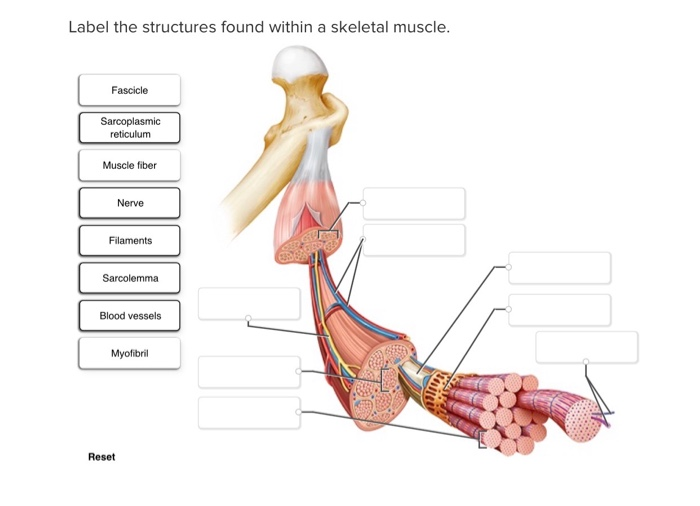
Solved Label the structures found within a skeletal muscle ...
Fetal Pig Dissection Manual Answer Booklet - City University of … ACTIVITY 10: For Part A, label structures A, B, C, and D on the image below. For Part B, complete the ... we have no conscious control over smooth muscle or cardiac muscle function, whereas we can consciously control to some extent all skeletal muscles. Can you consciously control your breathing? What does this tell you

Neurolemmocyte On Skeletal Muscle Model - Human Anatomy ...
Skeletal Muscle Histology Slide Identification and Labeled Diagram ... From the skeletal muscle histology slide, you might identify the following important structures under the light microscope. Please try to find out these structures from the skeletal muscle slide labeled images. #1. Longitudinal section of skeletal muscle #2. Cross-section of skeletal muscle #3. Skeletal muscle fibers of the longitudinal section #3.

The Multi-Scale, Three-Dimensional Nature of Skeletal Muscle ...
Solved Muscle Cell Label the structures of a skeletal muscle - Chegg Expert Answer 100% (16 ratings) 1) Sarcolemma 2) myofib … View the full answer Transcribed image text: Muscle Cell Label the structures of a skeletal muscle fiber. Nucleus Myofibril Sarcolemma Sarcoplasmic reticulum Openings into T tubules < Prev 3 of 15 !!! Next > Thinkinys - How to write a boty The Good Cre.

Label structure of skeletal muscle Diagram | Quizlet
Fluorescent Labeling - What You Should Know - PromoCell Improved fluorescent dyes are now enabling previously impossible studies of cellular structures and cellular processes. PromoCell provides a wide range of high-quality fluorophores for fluorescent labeling of diverse biomolecules: protein labeling, antibody labeling, nucleic acid labeling (DNA labeling, RNA labeling), as well as complete labeling kits and ready-to-use …

Muscle Structure (labelled), illustration - Stock Image ...
Solved Label the diagram below to review the basic structure - Chegg Label the diagram below to review the basic structure of skeletal muscle. Drag the labels into the boxes indicating the structure on the figure. Z line Myosin filament Nucleus eee A band O OC ES em Myofibril BASES ULD SEE Actin filament Sarcomere Sarcoplasmic reticulum I band Sarcolemma Sarcolemma
695 Deltoid Illustrations & Clip Art - iStock
Skeletal Muscle Labeling | Biology Quiz - Quizizz Skeletal Muscle Labeling DRAFT. 22 minutes ago. by ggard. Played 0 times. 0. 9th - 10th grade . Biology. 0% average accuracy. 0. Save. ... Q. Skeletal Muscle contraction is initiated when the _____ sends a message to the muscle cell. ... Microscopic Bone Structure . 1.6k plays . 19 Qs . Skeletal/Muscular Systems . 5.3k plays . Science - 5th ...

Organisation d'une fibre musculaire. Les fibres musculaires ...
11.2: Microscopic Anatomy of Skeletal Muscles - Biology LibreTexts Microscopic Anatomy of Skeletal Muscles. Skeletal muscle is found attached to bones. It consists of long multinucleate fibers. The fibers run the entire length of the muscle they come from and so are usually too long to have their ends visible when viewed under the microscope. The fibers are relatively wide and very long, but unbranched.
Skeletal Muscle Tissue Anatomy and Structure
The excitation–contraction coupling mechanism in skeletal muscle 24-01-2014 · The excitation–contraction coupling (ECC) mechanism in skeletal muscle. Ca 2+ cell homeostasis and signalling result from dynamic interactions between mechanisms that provoke an increase of cytoplasmic free Ca 2+ and those that reduce it. In the specific case of striated muscles, contraction and relaxation mechanisms are both regulated by rapid changes …
Skeletal muscle structure & function
Fluorescent Labeling - What You Should Know - PromoCell Definition Fluorescent labeling is the process of binding fluorescent dyes to functional groups contained in biomolecules so that they can be visualized by fluorescence imaging (nature.com). The availability of new fluorophores has dramatically changed the possibilities for the sensitive detection of biomolecules and the analysis of their interactions. Improved fluorescent dyes are now ...
Biology, Animal Structure and Function, The Musculoskeletal ...
10.2 Skeletal Muscle - Anatomy & Physiology These tissues include the skeletal muscle fibers, blood vessels, nerve fibers, and connective tissue. Each skeletal muscle has three layers of connective tissue that enclose it, provide structure to the muscle, and compartmentalize the muscle fibers within the muscle ( Figure 10.2.1 ).
Anatomy Review Skeletal Muscle Tissue - Anatomy Review ...
Label structure of skeletal muscle Diagram | Quizlet Label structure of skeletal muscle 4.0 5 Reviews STUDY Learn Write Test PLAY Match + − Created by danielaaaa04 Terms in this set (8) myofibrils ... sarcoplasmis reticulum ... sarcolemma ... epimysium ... perimysium ... endomysium ... fascicle ... muscle fiber ... Sets found in the same folder Sarcomere 11 terms MrDNorman TEACHER
Muscle-Skeletal Muscle-Gross and Ultra Structure - Online ...
Pathophysiology of Muscle Contractures in Cerebral Palsy Skeletal muscle is composed of bundles of muscle fibers called fascicles. Individual fibers in these fascicles consist of myofibrils, which are composed of the contractile proteins actin and myosin. Connective tissue, which surrounds the muscle at many levels, is organized into epimysium, surrounding the whole muscle; perimysium, surrounding ...
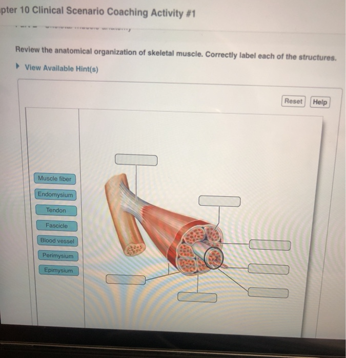
Solved apter 10 Clinical Scenario Coaching Activity #1 ...
SKELETAL MUSCLE ORGANIZATION - Brigham Young University-Idaho Each skeletal muscle cell, also called a muscle fiber, develops as many embryonic myocytes fused into one long, multi-nucleated skeletal muscle cell. These muscle fibers are bound together into bundles, or fascicles, and are supplied with a rich network of blood vessels and nerves. The fascicles are then bundled together to form the intact muscle.

Muscular System Functions & Organs | What Does the Muscular System Do? Video
Quiz: Structure of Skeletal Muscle - CliffsNotes Quiz: Connective Tissue Associated with Muscle Tissue; Structure of Skeletal Muscle; Quiz: Structure of Skeletal Muscle; Muscle Contraction; Types of Muscles; Quiz: Muscle Contraction; Muscle Metabolism; Structure of Cardiac and Smooth Muscle; Quiz: Structure of Cardiac and Smooth Muscle; The Muscular System. Quiz: Skeletal Muscle Actions ...
3,336 Muscle Cells Stock Photos, Pictures & Royalty-Free ...
PDF PowerPoint Presentation A motor neuron and all of the skeletal muscle cells it stimulates is called a (1) . The axon of each motor neuron has numerous endings called (2) . The actual gap between ... 14. Relative to general terminology concerning muscle activity, first label the following structures on Figure 6—5: insertion, origin, tendon, resting muscle, and ...

10.2 Skeletal Muscle – Anatomy & Physiology
PDF Biology 201: Muscles & Muscle Tissue 1) Label the structures of a ... Source Lesson: Muscle Fiber: Structures, Contraction & Relaxation 4) Label the structures in the diagram below. Some terms may be used more than once.

STRUCTURE OF A SKELETAL MUSCLE FIBER Diagram | Quizlet
Pathophysiology of Muscle Contractures in Cerebral Palsy However, when monoclonal antibodies were used to label different myosin heavy chain isoforms in the biceps brachii, a dramatic increase in percentage of type 2 fiber was reported, with type 2X myosin heavy chain fibers making up 30% of CP muscle compared with 4% of typically developing muscle. 46 Clearly variation exists among muscles, and further studies are needed …
Skeletal muscle - Wikipedia
108 Muscle Quizzes Online, Trivia, Questions & Answers - ProProfs 02-07-2022 · ProProfs, one of the popular quiz builder platforms, has more than 108 muscle quizzes which have already been played around 163954 times. Choose the game quiz you like and play as many times as you want

Skeletal muscle - Wikipedia
Skeletal Muscle: Definition, Function, Structure, Location | Biology ... Skeletal muscle, as the name implies, is any muscles that connects to and controls the motions of the skeleton. In all there are somewhere between 600 and 900 muscles in the human body, but an exact number is hard. Many muscles are obscurely small or are sometimes grouped together with similar muscles.

1,976 Skeletal Muscle Stock Photos, Pictures & Royalty-Free ...
Ultrastructure of Muscle - Skeletal - Sliding Filament - TeachMeAnatomy Skeletal - striated muscle that is under voluntary control from the somatic nervous system. Identifying features are cylindrical cells and multiple peripheral nuclei. Cardiac - striated muscle that is found only in the heart. Identifying features are single nuclei and the presence of intercalated discs between the cells.
Ch. 7
Drag each label to the correct location on the diagram. - Brainly.com 1. Muscle: soft, contractile tissue important to produce force and motion in animals. 2. Fascicle: multiple bundles of skeletal muscle fibres which is surrounded by a type of connective tissue called perimysium. 3. Muscle fibres: bundles of cylindrical organelles myofibrils formed by the fusion of myoblasts via myogenesis process. 4.
SKELETAL MUSCLE ORGANIZATION
Structure of Skeletal Muscle | SEER Training Each organ or muscle consists of skeletal muscle tissue, connective tissue, nerve tissue, and blood or vascular tissue. Skeletal muscles vary considerably in size, shape, and arrangement of fibers. They range from extremely tiny strands such as the stapedium muscle of the middle ear to large masses such as the muscles of the thigh.
Perbedaan Otot Lurik, Otot Polos, dan Otot Jantung
Naming Skeletal Muscles | Anatomy and Physiology I | | Course Hero The skeletal muscle's anatomical location or its relationship to a particular bone often determines its name. For example, the frontalis muscle is located on top of the frontal bone of the skull. Similarly, the shapes of some muscles are very distinctive and the names, such as orbicularis, reflect the shape.

A&P 1- CHAPTER 9 MASTERING ASSIGNMENTS Flashcards | Quizlet
Types of muscle cells: Characteristics, location, roles | Kenhub
SKELETAL MUSCLE ORGANIZATION

Muscle structure – muscle under the microscope — Science ...
12.3 Types of Muscle Tissue – Human Biology

Scheme of skeletal muscle and associated structures. (Left ...

10.2 Skeletal Muscle – Anatomy & Physiology
Muscle Structure Labeling (KEY)

11.4 Identify the skeletal muscles and give their origins ...
Post a Comment for "40 label the structures of a skeletal muscle"