44 dna labeling diagram
DNA labeling.pdf - DNA Labeling Nucleotide Pyrimidine... - Course Hero Leading strand Lagging strand Primase Ligase HelicaseOkazaki fragment Single strand binding proteinLabel the diagram%" ,DN)A polymerase adds nucleotides ± u001d' to °'² Replication fork is formed,DN)A polymerase attaches to the primerOkazaki fragments bound by ligase,DN)A helicase unwinds ,DN)ARearrange the steps to indicate the correct order%" DNA Sequencing: 7 Methods Used for DNA Sequencing The seven important methods used for DNA sequencing are: (1) Sanger’s Method (2) Maxam and Gilbert Method (3) Hybridization Method (4) Pal Nyren’s Method (5) Automatic DNA Sequencer (6) Slab Gel Sequencing Systems and (7) Capillary Gel Electrophoresis. DNA sequencing is the determination of the precise sequence of nucleotides in a sample of ...
Genetically Modified Organisms (GMOs) | Learn Science at Scitable … The pharmaceutical industry is another frontier for the use of GMOs. In 1986, human growth hormone was the first protein pharmaceutical made in plants (Barta et al., 1986), and in 1989, the first ...
Dna labeling diagram
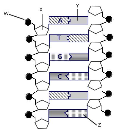
› Structure › MMDBMolecular Modeling Database (MMDB) Help Document In the illustration to the right, for example, the P53 tumor suppressor (accession 1TUP) is bound to double-stranded DNA, as viewed in the free Cn3D stand-alone program. The three-dimensional structure shows the functional shape of the protein and can be used to infer the specific amino acids that are active in binding to DNA. Achiever Papers - We help students improve their academic standing Professional academic writers. Our global writing staff includes experienced ENL & ESL academic writers in a variety of disciplines. This lets us find the most appropriate writer for any type of assignment. PDF Bell Ringer: DNA Diagram Labeling - Denton ISD Bell Ringer: DNA Diagram Labeling Identify the circled portions of the DNA Diagram and answer the 2 questions. A. _____ B. _____
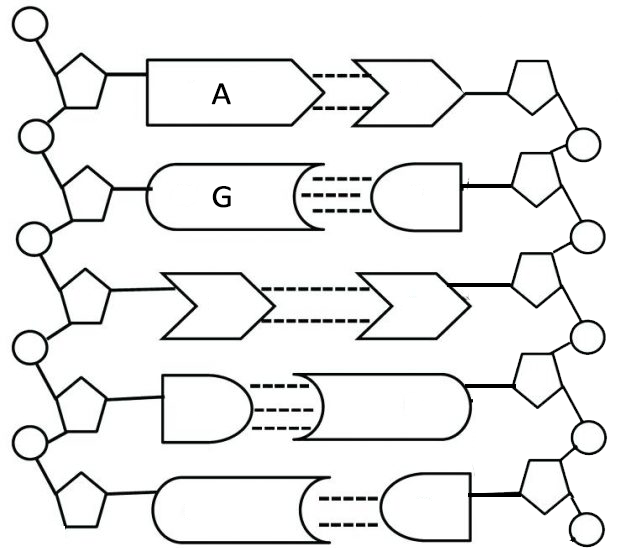
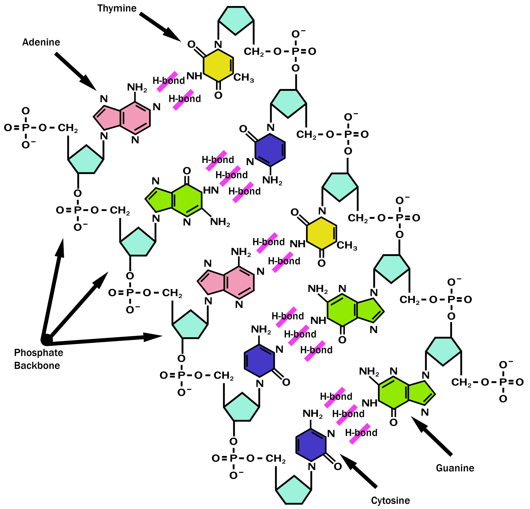
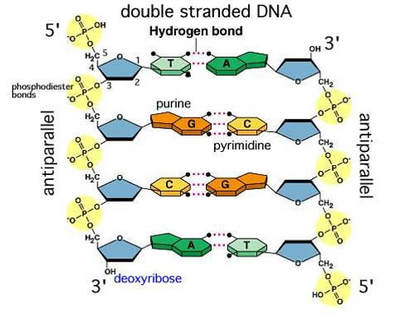
Dna labeling diagram. DNA_Structure_Labeling_Diagram_-instruction (1).pdf - DNA... DNA Structure Be able to label the following: 1. 5' end 2. 3' end 3. Phosphate (circled) 4. Deoxyribose (circled) 5. Purine 6. Pyrimidine 7. Deoxyribose carbon numbering 8. Hydrogen bond 9. Covalent bond 10. Adenine 11. Thymine 12. Guanine 13. Cytosine 14. Nucleotide Here's a blank diagram for you to practice your labeling! brd.nci.nih.gov › brd › sopLabeling SOP - National Institutes of Health Labeling SOP Page 4 of 22 Size: 1” W x 1” H BSI Report Generic_Lab_Label_Report / Standard DNA Label Report BradySoft Label Name Label: Lab/Standard Labels/ Standard 2D DNA Narrow Sample Label 1x1.lab Describer: DSC/Standard Labels/ Standardized DNA Label.dsc 4.5 Standard 2D Short 3 Across Label (.5” x 1” Label) 1. Dna labeling - Teaching resources - Wordwall Dna labeling Examples from our community 1465 results for 'dna labeling' DNA Labeling Labelled diagram by Azimmer Dna labeling Labelled diagram by Tclemmons G7 G9 Biology DNA Structure Labeling Labelled diagram by Mtaylor0182 G9 G10 Science DNA labeling Review Labelled diagram by Andrewtm G7 Science Community Items - Group 1 Open the box DNA Structure Labeling Diagram | Quizlet Start studying DNA Structure Labeling. Learn vocabulary, terms, and more with flashcards, games, and other study tools. 16 terms · double helix → the spiral-staircase structure…, nitrogen base → a subunit of a nucleotide in D…, pyrimidine → a nitrogen base that has a sin…, purine → a nitrogen base that has a dou…, thymine → ...
DNA Replication Labeling Diagram | Quizlet DNA Replication Labeling. STUDY. Learn. Flashcards. Write. Spell. Test. PLAY. Match. Gravity. Created by. gcpayton TEACHER. Terms in this set (11) Single Stranded Binding Protein. Binds to and stabilizes single-stranded DNA until it can be used as a template. Helicase. An enzyme that unwinds the DNA double helix during DNA replication. Dna diagram - Teaching resources - Wordwall Dna labeling Labelled diagram. by Tclemmons. G7 G9 Biology. DNA Labeling Labelled diagram. by Azimmer. Earth's Layers Diagram Labelled diagram. by Torkelson. Eastern USA Map Diagram Labelled diagram. by Jlozanos0003. G2 G3 G4 G5 G6 G7 G8 G9 G10 G11 G12 University Geography. Cellular Respiration Diagram Labelled diagram. Label DNA and Replication - Google Slides Label the diagram: DNA polymerase adds nucleotides (5' to 3') Replication fork is formed. DNA polymerase attaches to the primer. Okazaki fragments bound by ligase. DNA helicase unwinds DNA. Rearrange the steps to indicate the correct order: 1. Enzyme that unwinds DNA. Label the DNA molecule. Diagram | Quizlet Start studying Label the DNA molecule.. Learn vocabulary, terms, and more with flashcards, games, and other study tools.
Biotechnology - Wikipedia Biotechnology is the integration of natural sciences and engineering sciences in order to achieve the application of organisms, cells, parts thereof and molecular analogues for products and services. The term biotechnology was first used by Károly Ereky in 1919, meaning the production of products from raw materials with the aid of living organisms. Förster resonance energy transfer - Wikipedia Förster or fluorescence resonance energy transfer (FRET), resonance energy transfer (RET) or electronic energy transfer (EET) is a mechanism describing energy transfer between two light-sensitive molecules (chromophores). A donor chromophore, initially in its electronic excited state, may transfer energy to an acceptor chromophore through nonradiative dipole–dipole coupling. DNA Molecule Label Diagram | Quizlet Molecule found on the side of a DNA molecule Double Helix two strands of nucleotides wound about each other; structure of DNA Thymine the nucleotide that hydrogen bonds with the nucleotide adenine in DNA Adenine the nucleotide that hydrogen bonds with the nucleotide thymine in DNA or with uracil in RNA Guamine PHSchool.com Retirement–Prentice Hall–Savvas Learning Company PHSchool.com was retired due to Adobe’s decision to stop supporting Flash in 2020. Please contact Savvas Learning Company for product support.
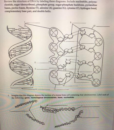
Review the Structure of DNA
en.wikipedia.org › wiki › BiotechnologyBiotechnology - Wikipedia Pharmacogenomics (a combination of pharmacology and genomics) is the technology that analyses how genetic makeup affects an individual's response to drugs. Researchers in the field investigate the influence of genetic variation on drug responses in patients by correlating gene expression or single-nucleotide polymorphisms with a drug's efficacy or toxicity.
DNA Structure — Overview & Diagrams - Expii
DNA Replication Labeling Diagram | Quizlet The primer synthesized by primase enzyme DNA Polymerase on Leading Strand synthesizes new DNA only in the 5' to 3' direction Ligase An enzyme that connects two fragments of DNA to make a single fragment Lagging Strand The strand in replication that is copied 3' to 5' as Okazaki fragments and then joined up. DNA Polymerase on lagging strand

DNA Structure Labeling Diagram | Quizlet
Transcription vs Translation - Difference and Comparison | Diffen For Transcription, RT-PCR, DNA microarray, In-situ hybridization, Northern blot, RNA-Seq is quite often used for measurement and detection. For Translation, western blotting, immunoblotting, enzyme assay, Protein sequencing, Metabolic labeling, proteomics is …
File:DNA-labels.png - Wikimedia Commons
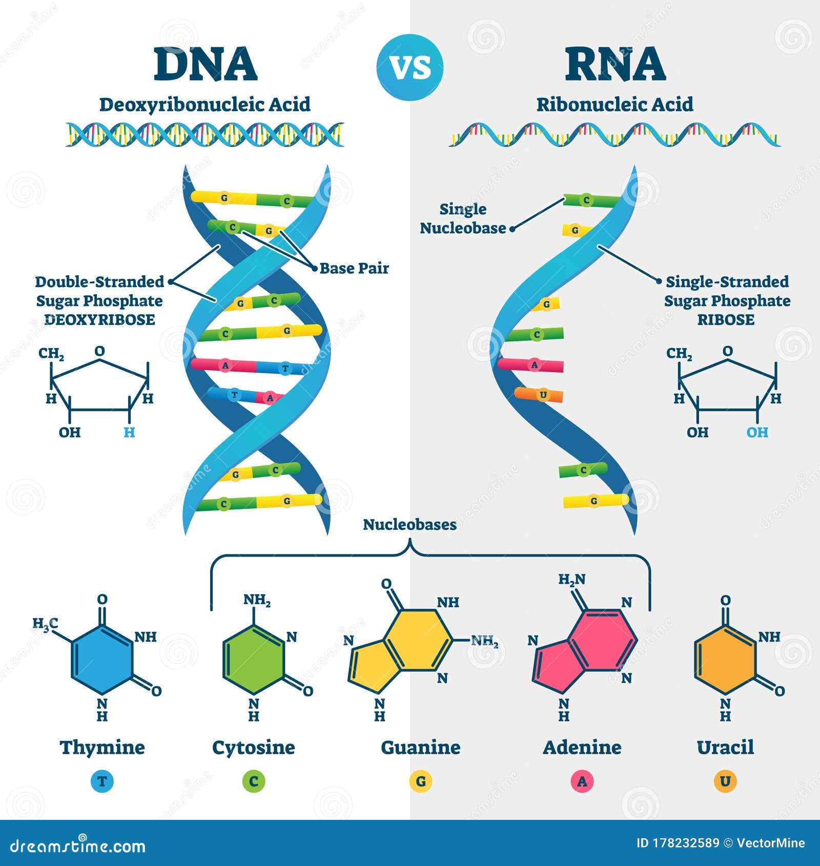
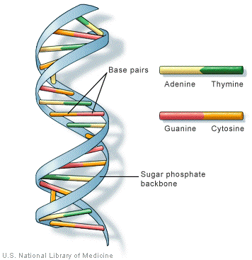
Nucleotide Structure: DNA Diagram | Science Trends The structure of DNA and RNA is also different. DNA is known for its double helix structure. The double helix is two strands that are intertwined with one another thanks to the complementary bases. RNA is a single-stranded molecule by contrast. The double helix form of DNA helps keep the genetic code intact.

DNA Model Labeling Diagram | Quizlet
en.wikipedia.org › wiki › IronIron - Wikipedia For U.S. food and dietary supplement labeling purposes the amount in a serving is expressed as a percent of Daily Value (%DV). For iron labeling purposes 100% of the Daily Value was 18 mg, and as of May 27, 2016 [update] remained unchanged at 18 mg. [166] [167] A table of the old and new adult daily values is provided at Reference Daily Intake .

Diagram Of A DNA Structure || Labelled Diagram Of DNA ...
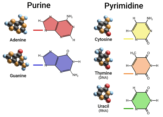
DNA Replication: Process with Diagrams - Turito Step 1: Formation of Replication Forks. Before DNA can be reproduced, it must first be "unzipped" into two single strands. Adenine (A), cytosine (C), thymine (T), and guanine (G) are the four nucleotides that form pairings between the two strands of DNA. Adenine only binds to thymine, while cytosine only binds to guanine.
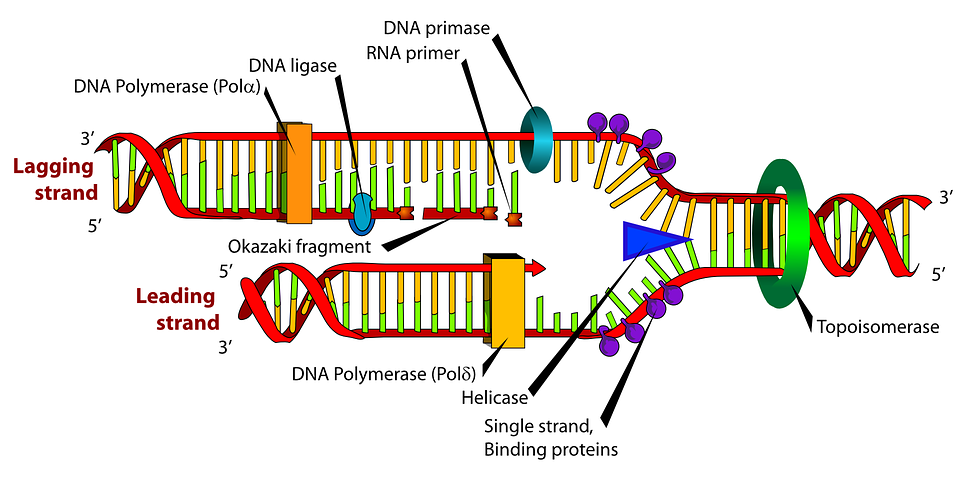
Diagram Dna Biology - Free vector graphic on Pixabay
achieverpapers.comAchiever Papers - We help students improve their academic ... Professional academic writers. Our global writing staff includes experienced ENL & ESL academic writers in a variety of disciplines. This lets us find the most appropriate writer for any type of assignment.
Notes: DNA Structure Topic ppt download
› indexPHSchool.com Retirement–Prentice Hall–Savvas Learning Company PHSchool.com was retired due to Adobe’s decision to stop supporting Flash in 2020. Please contact Savvas Learning Company for product support.

DNA function & structure (with diagram) (article) | Khan Academy
DNA Structure labeled diagram Diagram | Quizlet DNA Structure labeled diagram 4.8 (4 reviews) + − Flashcards Learn Test Match Created by study-14 Terms in this set (7) 1 nucleotide sugar molecule phosphate group and 1 nitrogen base Sugar molecule ... Phosphate group ... sugar phosphate backbone ... nitrogen bases adenine, thymine, cytosine and guanine

A) Strategy for terminal or internal DNA labeling using ...
Dna labeling - Labelled diagram - Wordwall Dna labeling - Labelled diagram. Sugar (Deoxyribose), phosphate , nitrogen base Adenine, nitrogen base Guanine, Hydrogen bond, nitrogen base cytosine, nitrogen base thymine, backbone.
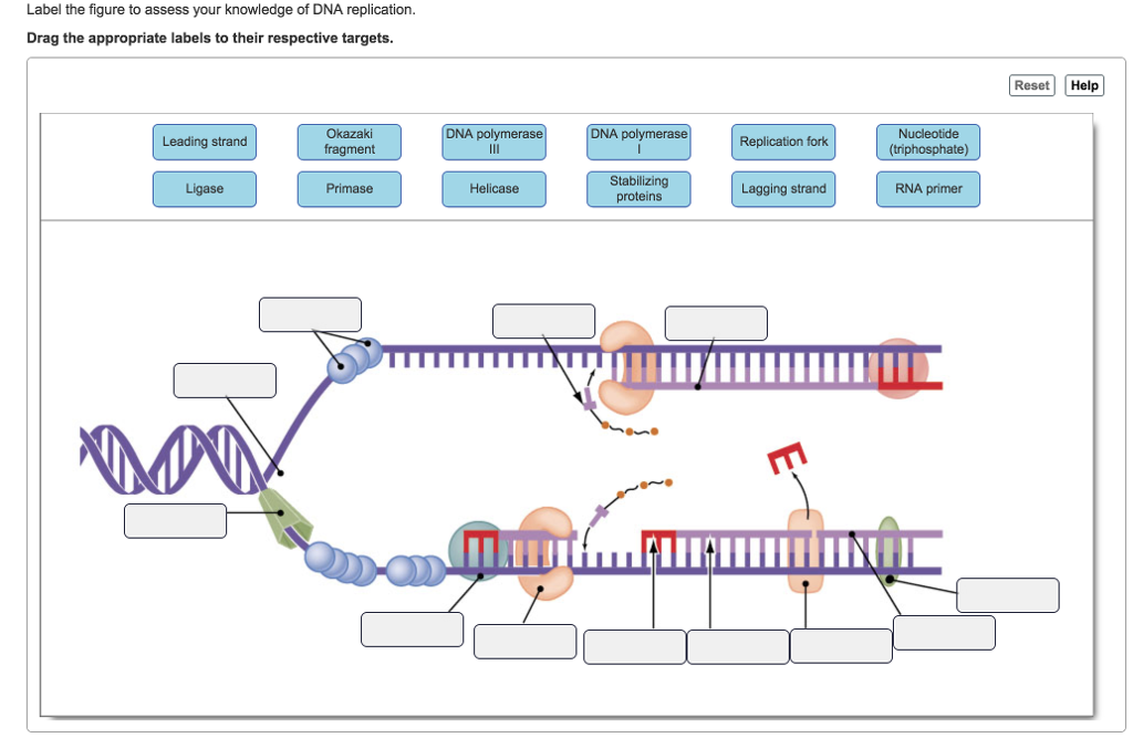
Answered: Label the figure to assess your… | bartleby
› scitable › topicpageGenetically Modified Organisms (GMOs) | Learn Science at Scitable The pharmaceutical industry is another frontier for the use of GMOs. In 1986, human growth hormone was the first protein pharmaceutical made in plants (Barta et al., 1986), and in 1989, the first ...

DNA Labeling Diagram | Quizlet
Molecular Modeling Database (MMDB) Help Document Source Database. The Molecular Modeling DataBase (MMDB) is a database of experimentally determined three-dimensional biomolecular structures, and is also referred to as the Entrez Structure database. It is a subset of three-dimensional structures obtained from the RCSB Protein Data Bank (PDB), excluding theoretical models.The data processing procedure at NCBI results …

A) Concept of metabolic DNA labeling. Deoxynucleosides with ...
DNA function & structure (with diagram) (article) | Khan Academy DNA function & structure (with diagram) (article) | Khan Academy DNA structure and function DNA is the information molecule. It stores instructions for making other large molecules, called proteins. These instructions are stored inside each of your cells, distributed among 46 long structures called chromosomes.
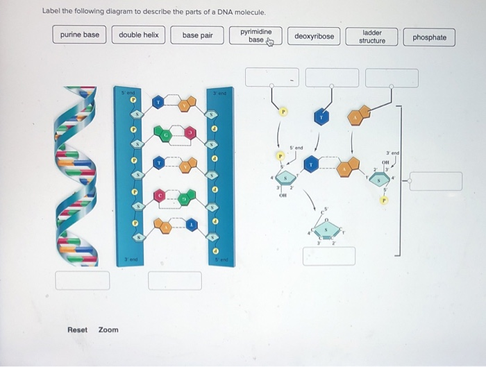
Solved Label the following diagram to describe the parts of ...
Labeling SOP - National Institutes of Health Labeling SOP Page 4 of 22 Size: 1” W x 1” H BSI Report Generic_Lab_Label_Report / Standard DNA Label Report BradySoft Label Name Label: Lab/Standard Labels/ Standard 2D DNA Narrow Sample Label 1x1.lab Describer: DSC/Standard Labels/ Standardized DNA Label.dsc 4.5 Standard 2D Short 3 Across Label (.5” x 1” Label) 1.

Image: DNA Structure
Biology DNA Labeling Diagram | Quizlet Start studying Biology DNA Labeling. Learn vocabulary, terms, and more with flashcards, games, and other study tools.

a Immuno-PCR using site-specific DNA labeling and RCA ...
DNA Labeling: Overview & Applications DNA labeling is a technique used in molecular labs to detect and purify DNA molecules. It's done by the introduction of a fluorescent molecule per DNA strand and a quantitative fragmentation of the DNA, followed by the incorporation of one label per DNA fragment.

DNA Replication Labelling Diagram | Quizlet
Iron - Wikipedia Iron (/ ˈ aɪ ər n /) is a chemical element with symbol Fe (from Latin: ferrum) and atomic number 26. It is a metal that belongs to the first transition series and group 8 of the periodic table.It is, by mass, the most common element on Earth, right in front of oxygen (32.1% and 30.1%, respectively), forming much of Earth's outer and inner core.It is the fourth most common …
Scheme Deoxyribonucleic Stock Illustrations – 17 Scheme ...
DNA: Structure, Forms and Functions (With Diagram) - Biology Discussion DNA or Deoxyribonucleic acid is a type of nucleic acid. It is present in all living cells of bacteria, trees, dogs, cats and human. Some viruses also contain DNA. DNA was discovered in 1868 by the German biochemist, Friedrich Miescher who called it nuclein.
Set of Molecule Badge. Connected Hexagonal Molecule Badge ...
DNA_Labeling_diagram (1).pdf - | Course Hero View DNA_Labeling_diagram (1).pdf from BIOL BIOL301 at American Public University.

DNA function & structure (with diagram) (article) | Khan Academy
PDF Bell Ringer: DNA Diagram Labeling - Denton ISD Bell Ringer: DNA Diagram Labeling Identify the circled portions of the DNA Diagram and answer the 2 questions. A. _____ B. _____

Fluorescent Labeling of Plasmid DNA and mRNA: Gains and ...
Achiever Papers - We help students improve their academic standing Professional academic writers. Our global writing staff includes experienced ENL & ESL academic writers in a variety of disciplines. This lets us find the most appropriate writer for any type of assignment.

Sequence‐specific Methyltransferase‐Induced Labeling of DNA ...
› Structure › MMDBMolecular Modeling Database (MMDB) Help Document In the illustration to the right, for example, the P53 tumor suppressor (accession 1TUP) is bound to double-stranded DNA, as viewed in the free Cn3D stand-alone program. The three-dimensional structure shows the functional shape of the protein and can be used to infer the specific amino acids that are active in binding to DNA.

The Structure of DNA

Visual Learning
Covalent labeling of nucleic acids - Chemical Society Reviews ...

Metabolic DNA labeling using alkyne- or alkene-modified ...
Solved Review the structure of DNA by labeling these | Chegg.com

Schematic illustration of the detection strategy and chemical ...
Redox coding of DNA bases: structures of redox-labelled dNTPs ...

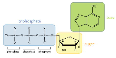
Identifying the Components of DNA

A) Schematic overview over the dual labeling of DNA using P1 ...

Label the parts of the DNA in the diagram given below ...
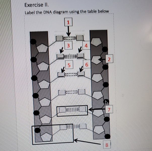
Solved Exercise II. Label the DNA diagram using the table ...
Solved] Worksheet - Structure of DNA and Replication ...
Life Sciences Cyberbridge
2.6 DNA & RNA Structure

Unnatural base pairs for enzymatic site-specific labeling of ...

7.1 DNA Structure - SL/HL2 Biology Ferguson

Biology CSF Chapter 14 HW Flashcards | Quizlet
DNA - structure

DNA Labeling by Ligand Inducible Secondary Structure - Peng ...
Label the parts of the DNA in the diagram given below ...
Review Guide: DNA and Genetic Engineering
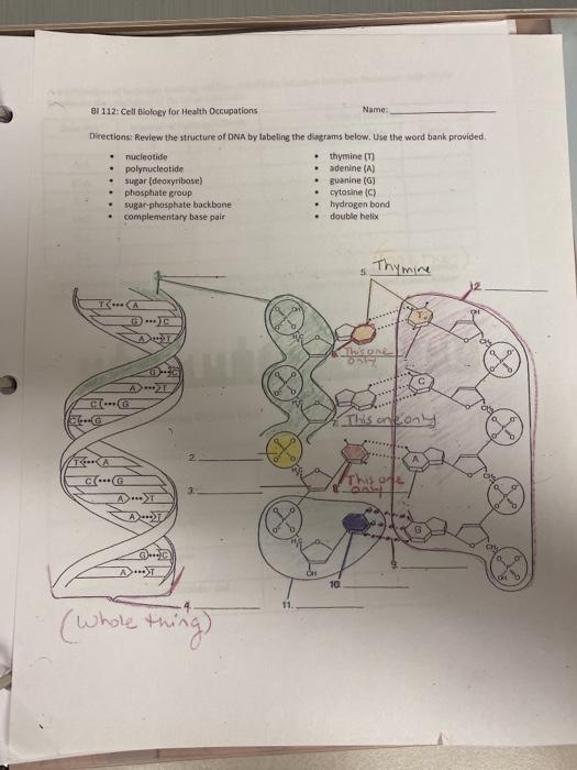
Solved BI 112: Cell Biology for Health Occupations Name ...
Post a Comment for "44 dna labeling diagram"