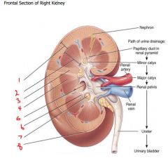
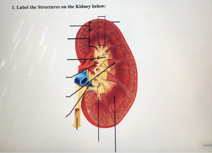
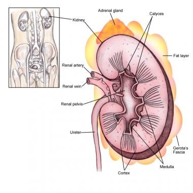
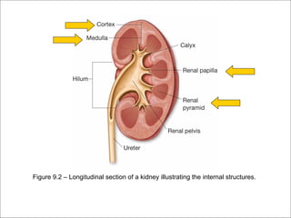
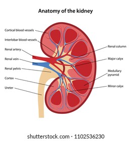
38 label the structures of the kidney
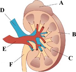
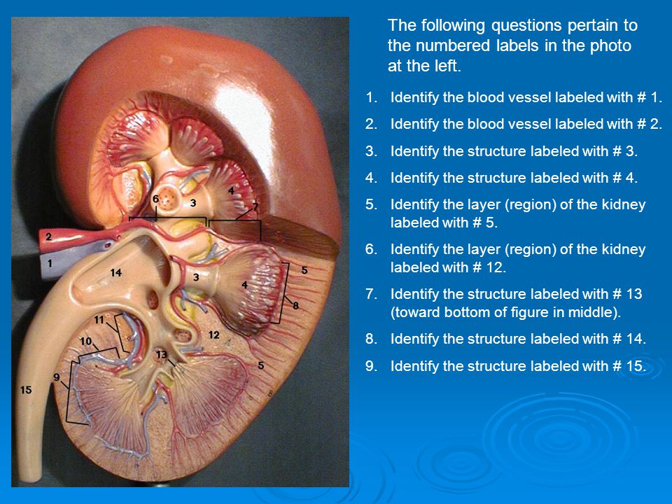
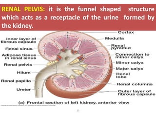
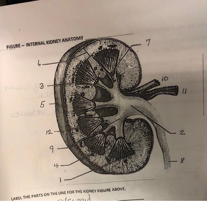
Label the kidney Quiz - PurposeGames.com This is an online quiz called Label the kidney. There is a printable worksheet available for download here so you can take the quiz with pen and paper. Your Skills & Rank. Total Points. 0. Get started! Today's Rank--0. Today 's Points. One of us! Game Points. 9. You need to get 100% to score the 9 points available. Solved Label the structures of the kidney. Renal Pelvis - Chegg The anatomy of kidney shows two regions. The outer region is called renal cortex and the inner dark r … View the full answer Transcribed image text: Label the structures of the kidney. Renal Pelvis Major Calyx Renal Cortex Minor Calyx Ureter Major Calyx Renal Cortex Renal Pelvis Minor Calyx Ureter Renal Pyramid Renal Medulla
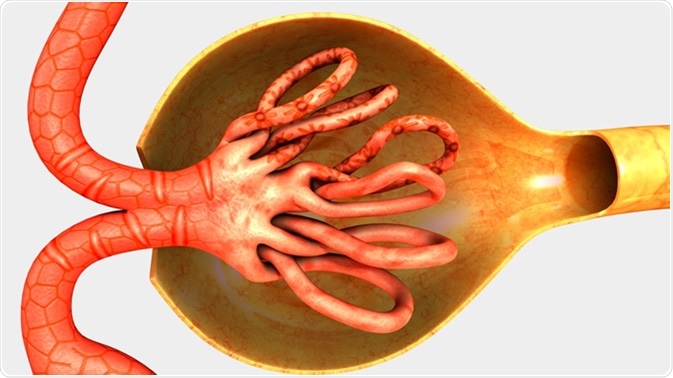
Urinary System - Label the Kidney and Nephron - The Biology Corner Students drag labels to the structures on the slide. Also, the diagram shows the relationship between the aorta, vena cava, and the renal vessels. While these aren't part of the urinary system, they are important in the physiology of the kidney. On the second slide, viewers see a close-up of a kidney that's been cut to show the internal structures.

Label the structures of the kidney
A&P 139 Urinary System Flashcards | Quizlet Label the external anatomy of the kidney, using the hints provided. Place the following vessels in the correct order of blood flow, starting with the vessel that is a branch off the aorta. Place the following structures found in the female pelvis is order from posterior to anterior. collepals.com › orders › studOrder Panel - collepals.com - College Pal × Are you sure? NO YES YES Kidney-Structure, Anatomy and Function - Online Biology Notes Kidney-Structure, Anatomy and Function Gross Structure Kidneys are bean-shaped organs, about 11 cm long, 6 cm wide, 3 cm thick and weigh 150 g. They are embedded in, and held in position by, a mass of adipose tissue. Each kidney is enclosed by a thin tough fibrous connective tissue called renal capsule that protects it from infections and injuries.
Label the structures of the kidney. Ch. 17 Urinary System (Kidney Labeling) Quiz - PurposeGames.com This online quiz is called Ch. 17 Urinary System (Kidney Labeling) anatomy. This online quiz is called Ch. 17 Urinary System (Kidney Labeling) anatomy. English en. Login. Login Register Free Help; Start; Explore. Games; Playlists; Tournaments; Tags; The Wall; Badges; Leaderboard; Create. Create a Quiz; Create a Group; Create a Playlist; Groups. Structure of the Kidney (With Diagram) | Organs | Human Physiology Structure:. The kidneys have been bean-shaped appearance. The outer edge of the kidney is convex and the inner concave. Under a microscope a thin section of the kidney is seen to be composed of the following parts:. (A) Malpighian Body:. It is found in the cortex of the kidney. Kidney is composed ... › news › kidneyCareWatch out for Your Kidneys When You Use Medicines for Pain Aug 12, 2014 · Kidney damage happens because high doses of the drugs have a harmful effect on kidney tissue and structures. These drugs can also reduce the blood flow to the kidney. If you are older, your kidneys may have a stronger reaction to these medicines and you may need a smaller dose. Kidney disease from pain medicines is often preventable. Describe the structure of a human kidney with the help of a labelled ... The human kidney is a reddish-brown bean-shaped structure situated between the last thoracic and third lumbar vertebra close to the dorsal inner wall of the abdominal cavity. The kidney is covered by fibrous connective tissue, the renal capsule, which protects the kidney Internally, It consists of the outer dark cortex and an inner light ...
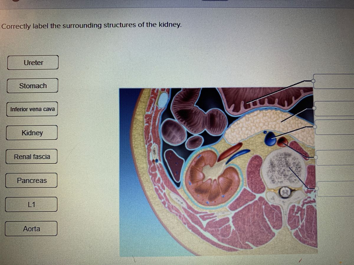
Ch. 25 Introduction - Anatomy and Physiology | OpenStax Label structures of the urinary system; Characterize the roles of each of the parts of the urinary system; ... (EPO) produced to stimulate red blood cell production is produced in the kidneys. The kidneys also perform the final synthesis step of vitamin D production, converting calcidiol to calcitriol, the active form of vitamin D. ... Correctly label the surrounding structures of the kidney. Correctly label the surrounding structures of the kidney. STUDY Learn Write Test PLAY Match Created by Sarah_Branning Terms in this set (5) Spleen ... Small intestine ... Colon ... Peritoneum ... Lumbar muscles ... The Structure and Function of the Kidneys - Verywell Health They normally range in size from 8 to 14 centimeters (or 3 to 5.5 inches). Each kidney weighs between 120 grams (about a quarter-pound) to 170 grams (0.4 lbs). These numbers vary based on a person's size, and abnormal-sized kidneys could be a sign of kidney disease. About 380 gallons (1,440 liters) of blood flow through the kidneys every day. Kidney Structure and Kidney Function Information The kidneys are a pair of bean shaped organs. In adults, a kidney is about 10 cm long, 6 cm wide and 4 cm thick. Each kidney weighs approximately 150-170 grams. Urine formed in the kidneys flow down to urinary bladder and then through the ureters. Each ureter is about 25 cm long and is a hollow tube- like structure made up of special muscles.
veux-veux-pas.fr › en › classified-adsAll classifieds - Veux-Veux-Pas, free classified ads Website All classifieds - Veux-Veux-Pas, free classified ads Website. Come and visit our site, already thousands of classified ads await you ... What are you waiting for? It's easy to use, no lengthy sign-ups, and 100% free! If you have many products or ads, create your own online store (e-commerce shop) and conveniently group all your classified ads in your shop! Webmasters, you can add your site in ... A&P 139 Urinary System Flashcards | Quizlet Label the structures of the renal corpuscle and juxtaglomerular apparatus. Label the CT scan of the abdominal contents using the hints provided. urethra maintaining constant GFR. Autoregulation refers to Label the internal anatomy of the kidney using the hints provided. emedicine.medscape.com › article › 117853-overviewType 2 Diabetes Mellitus: Practice Essentials, Background ... May 31, 2022 · Davies M, Heller S, Sreenan S, Sapin H, Adetunji O, Tahbaz A, et al. Once-Weekly Exenatide Versus Once- or Twice-Daily Insulin Detemir: Randomized, open-label, clinical trial of efficacy and safety in patients with type 2 diabetes treated with metformin alone or in combination with sulfonylureas. Diabetes Care. 2012 Dec 28. [QxMD MEDLINE Link]. › x-raysX-rays - what are they and different types | healthdirect Patients with kidney problems face greater risks when having contrast medium than other people. If you have kidney problems and need an x-ray with contrast medium, talk to your doctor first. Some people have an allergic reaction to iodine-containing contrast dye. Reactions can be mild, moderate or severe.
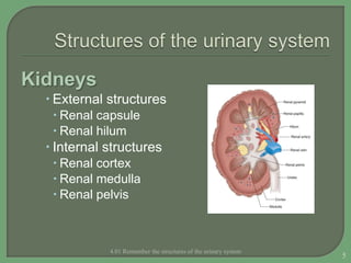
Kidney Anatomy and Function - Health Pages The kidneys are highly vascular (contain a lot of blood vessels) and are divided into three main regions: the renal cortex (outer region which contains about 1.25 million renal tubules), renal medulla (middle region which acts as a collecting chamber), and renal pelvis (inner region which receives urine through the major calyces).
Labeled Diagram of the Human Kidney - Bodytomy The vital structural components of a kidney are enclosed in a smooth but tough fibrous capsule called renal capsule. Inside this capsule, two distinct regions can be observed: a pale outer region called renal cortex, and a dark inner... The renal medulla comprises a set of 8-18 conical structures ...
Page: Journal of Investigative Dermatology Rademacher et al. show that the serine protease Esp from the abundant skin commensal Staphylococcus epidermidis processes pro‒IL-1β to mature, biologically active IL-1β produced by epidermal keratinocytes in the absence of host canonical processing by the inflammasome and caspase-1.
Kidneys: Anatomy, function and internal structure | Kenhub The posterior surfaces of both kidneys are related to certain neurovascular structures and muscles: 1 Artery: subcostal artery 2 Bones: 11th and 12th ribs 3 Nerves: subcostal, iliohypogastric, and ilioinguinal nerves 4 Muscles: diaphragm, psoas major, quadratus lumborum, transversus abdominis
Kidney Structures and Functions Explained (with Picture and Video ... Kidney Structure. The bean-shaped kidneys have an outer convex side and an inner concave side ...
go.drugbank.com › drugs › DB00264Metoprolol: Uses, Interactions, Mechanism of Action ... Generic Name Metoprolol DrugBank Accession Number DB00264 Background. Metoprolol is a selective beta-1 blocker commonly employed as the succinate and tartrate derivatives depending if the formulation is designed to be of immediate release or extended release. 2,9 The possibility of the generation of these formulations comes from the lower systemic bioavailability of the succinate derivative. 5 ...
A&P II (Ch 24 - 25) Flashcards | Quizlet nephron consists of. renal corpuscle and renal tubule. T/F: The juxtaglomerular apparatus is a structure of the nephron where the DCT contacts the afferent arteriole. true. Label the structures of a nephron in the figure. Correctly label the following components of the urinary system.
25.3 Gross Anatomy of the Kidney - OpenStax External Anatomy. The left kidney is located at about the T12 to L3 vertebrae, whereas the right is lower due to slight displacement by the liver. Upper portions of the kidneys are somewhat protected by the eleventh and twelfth ribs ( Figure 25.7 ). Each kidney weighs about 125-175 g in males and 115-155 g in females.
Labeled diagram of the human kidney royalty-free images - Shutterstock 189 labeled diagram of the human kidney stock photos, vectors, and illustrations are available royalty-free. See labeled diagram of the human kidney stock video clips. Image type.
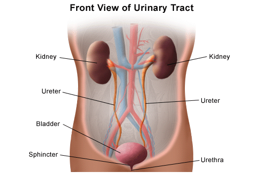
Urinary System Structures - Visible Body The kidneys, ureters, bladder, and urethra are the primary structures of the urinary system. They filter blood and remove waste from the body in the form of urine. The size and position of lower urinary structures vary with male and female anatomy. 1. Kidneys Filter Blood at the Top of the Urinary System.
Kidneys: Location, function, anatomy, pictures, and related diseases Inside the kidneys are a number of pyramid-shaped lobes. Each consists of an outer renal cortex and an inner renal medulla. Nephrons flow between these sections. Each nephron includes a filter,...
Kidneys: Anatomy, Location, and Function - Verywell Health Anatomy. Each person has two kidneys. The kidneys are located on either side of the spine, with the top of each kidney beginning around the 11th or 12th rib space. The kidneys are sandwiched between the diaphragm and the intestines, closer to the back side of the abdomen. Roughly the size of a closed fist, each kidney measures about 10 to 12 ...
Kidney Anatomy Labeling Diagram | Quizlet Kidney Anatomy Labeling 5.0 (3 reviews) + − Learn Test Match Created by lizhuver Terms in this set (11) Renal cortex ... Renal medulla ... Major calyx ... Papilla of Pyramid ... Renal pelvis ... Minor calyx ... Ureter ... Renal pyramid in renal medulla ... Renal column ... Fibrous capsule ... Renal hilum ... Urinary System - KIDNEY FOCUS
Kidney: Function and Anatomy, Diagram, Conditions, and ... - Healthline The kidneys are two bean-shaped organs in the renal system. They help the body pass waste as urine. They also help filter blood before sending it back to the heart. The kidneys perform many ...
Parts of the Kidney: Internal Anatomy of the Kidney - Study.com There are three major parts of the kidney: The renal cortex is the outer part of the kidney where blood is filtered. The renal medulla is the inner portion of the kidney where urine is formed. The ...
Kidney-Structure, Anatomy and Function - Online Biology Notes Kidney-Structure, Anatomy and Function Gross Structure Kidneys are bean-shaped organs, about 11 cm long, 6 cm wide, 3 cm thick and weigh 150 g. They are embedded in, and held in position by, a mass of adipose tissue. Each kidney is enclosed by a thin tough fibrous connective tissue called renal capsule that protects it from infections and injuries.
collepals.com › orders › studOrder Panel - collepals.com - College Pal × Are you sure? NO YES YES
A&P 139 Urinary System Flashcards | Quizlet Label the external anatomy of the kidney, using the hints provided. Place the following vessels in the correct order of blood flow, starting with the vessel that is a branch off the aorta. Place the following structures found in the female pelvis is order from posterior to anterior.
:watermark(/images/watermark_only_sm.png,0,0,0):watermark(/images/logo_url_sm.png,-10,-10,0):format(jpeg)/images/anatomy_term/collecting-duct-2/3Ge98cbJXXRsU4MT1NzJw_15Collecting_duct_of_kidney.png)


:watermark(/images/watermark_5000_10percent.png,0,0,0):watermark(/images/logo_url.png,-10,-10,0):format(jpeg)/images/overview_image/684/UScWzmJPRXIEJxzg40iQQ_kidneys_english.jpg)


:watermark(/images/watermark_only_sm.png,0,0,0):watermark(/images/logo_url_sm.png,-10,-10,0):format(jpeg)/images/anatomy_term/renal-cortex-2/pKVs9Ww61oXj7OfwTe3Tw_Cortex_renalis_02.png)




:background_color(FFFFFF):format(jpeg)/images/library/6707/minor_calyx.png)





Post a Comment for "38 label the structures of the kidney"