44 microscope label diagram
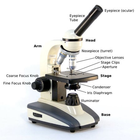
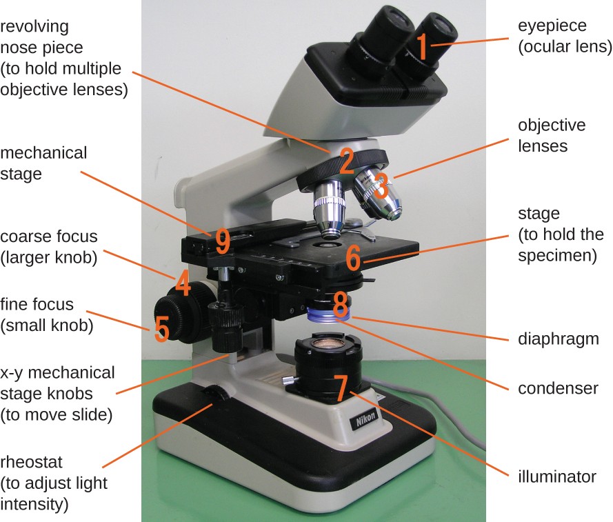
Interactive Eukaryotic Cell Model - CELLS alive WebSecretory Vesicle: Cell secretions - e.g. hormones, neurotransmitters - are packaged in secretory vesicles at the Golgi apparatus.The secretory vesicles are then transported to the cell surface for release. Cell Membrane: Every cell is enclosed in a membrane, a double layer of phospholipids (lipid bilayer).The exposed heads of the bilayer are "hydrophilic" … Parts of the Microscope with Labeling (also Free Printouts) A microscope is one of the invaluable tools in the laboratory setting. It is used to observe things that cannot be seen by the naked eye. Table of Contents 1. Eyepiece 2. Body tube/Head 3. Turret/Nose piece 4. Objective lenses 5. Knobs (fine and coarse) 6. Stage and stage clips 7. Aperture 9. Condenser 10. Condenser focus knob 11. Iris diaphragm
News: Breaking stories & updates - The Telegraph WebLatest breaking news, including politics, crime and celebrity. Find stories, updates and expert opinion.
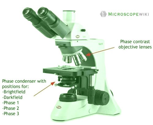
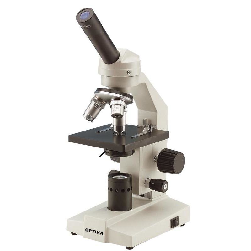
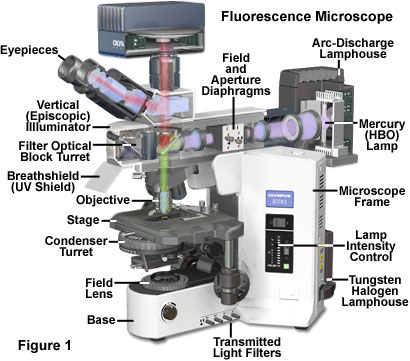
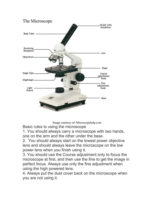
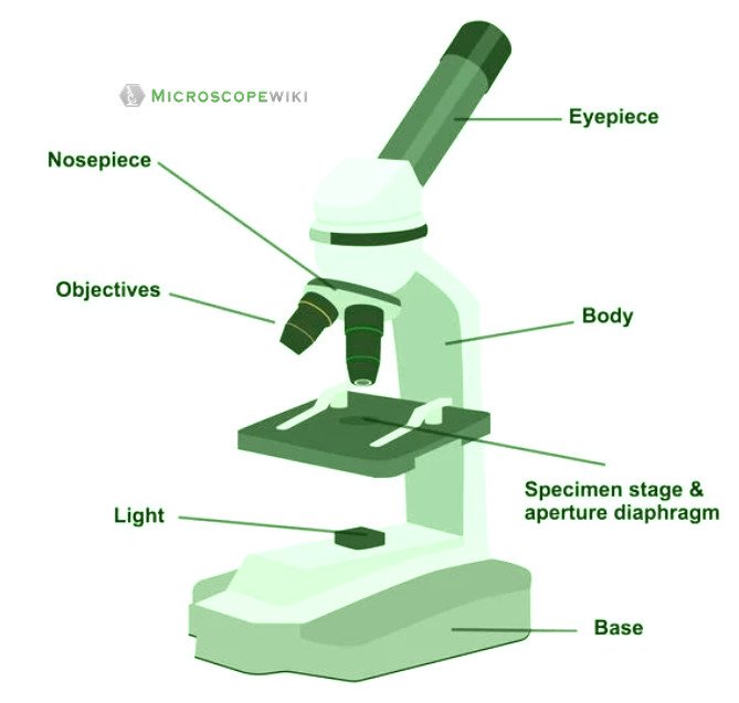
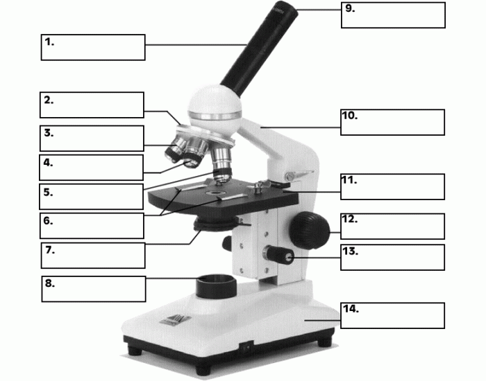
Microscope label diagram
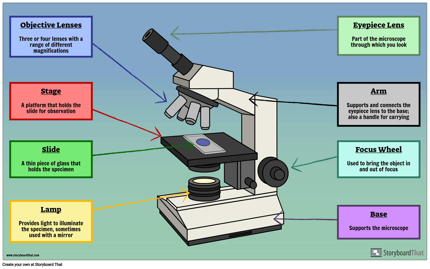
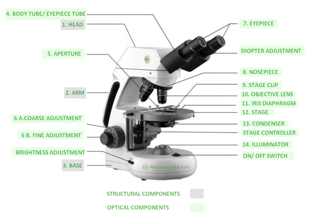
Microscope: Parts Of A Microscope With Functions And Labeled Diagram. Figure: A diagram of a microscope's components The microscope has three basic components: the head, the base, and the arm. Head:Occasionally, the head is considered the body. It holds the optical components of the upper part of the microscope. Base:The microscope's base provides great support. It is also equipped with miniature illuminators. Microscope Labeling Diagram | Quizlet Hold the slide in place on the stage. Nosepiece Holds the objective lenses and allows the lenses to rotate for viewing. Stage Supports the slide where the specimen is being viewed. Lamp Projects or reflects light upward through the diaphragm. Base Supports and stabilizes the microscope. Diaphragm Diagram of a Compound Microscope - Biology Discussion 1. It is noted first that which objective lens is in use on the microscope. 2. Stage micrometer is positioned in such a way that it is in the field of view. 3. The eyepiece is rotated so that the two scales, the eyepiece or ocular scale and the stage micrometer scale, are parallel. 4.
Microscope label diagram. Microscope Parts and Functions Microscope Parts and Functions With Labeled Diagram and Functions How does a Compound Microscope Work?. Before exploring microscope parts and functions, you should probably understand that the compound light microscope is more complicated than just a microscope with more than one lens.. First, the purpose of a microscope is to magnify a small object or to magnify the fine details of a larger ... Cell (biology) - Wikipedia WebCell shapes. Cell shape, also called cell morphology, has been hypothesized to form from the arrangement and movement of the cytoskeleton. Many advancements in the study of cell morphology come from studying simple bacteria such as Staphylococcus aureus, E. coli, and B. subtilis. Different cell shapes have been found and described, but how and why … Parts of Stereo Microscope (Dissecting microscope) – labeled diagram ... WebUnlike a compound microscope that offers a flat image, stereo microscopes give the viewer a 3-dimensional image that you can see the texture of a larger specimen. [In this image] Examples of Stereo & Dissecting microscopes. Major microscope brands (Zeiss, Olympus, Nikon, Amscope, Omano, Leica …) all produce stereomicroscopes. Videos | HowStuffWorks WebHowStuffWorks explains thousands of topics, from engines to lock-picking to ESP, with video and illustrations so you can learn how everything works.
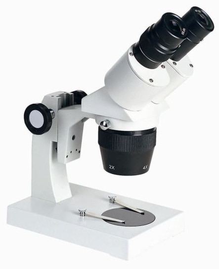
rsscience.com › stereo-microscopeParts of Stereo Microscope (Dissecting microscope) – labeled ... Labeled part diagram of a stereo microscope Major structural parts of a stereo microscope. There are three major structural parts of a stereo microscope. The viewing Head includes the upper part of the microscope, which houses the most critical optical components, including the eyepiece, objective lens, and light source of the microscope. Parts of a microscope with labeled diagram and functions Structural parts of a microscope: There are three major structural parts of a microscope. The head comprises the top portion of the microscope, which contains the most important optical components, and the eyepiece tube. The base serves as the microscope's support and holds the illuminator. The arm is the component of the microscope that ... rsscience.com › compound-microscope-parts-labeledCompound Microscope Parts – Labeled Diagram and their ... Labeled diagram of a compound microscope Major structural parts of a compound microscope Optical components of a compound microscope Eyepiece Eyepiece tube Objective lenses Nosepiece Specimen stage Coarse and fine focus knobs Rack stop Illuminator Condenser Abbe condenser Iris Diaphragm Condenser Focus Knob Summary An overview of microscopes Label the microscope — Science Learning Hub Web8. Juni 2018 · All microscopes share features in common. In this interactive, you can label the different parts of a microscope. Use this with the Microscope parts activity to help students identify and label the main parts of a microscope and then describe their functions.. Drag and drop the text labels onto the microscope diagram. If you want to …
Labeling the Parts of the Microscope | Microscope World Resources Labeling the Parts of the Microscope This activity has been designed for use in homes and schools. Each microscope layout (both blank and the version with answers) are available as PDF downloads. You can view a more in-depth review of each part of the microscope here. Download the Label the Parts of the Microscope PDF printable version here. Tongue Under Microscope with Labeled Diagram - AnatomyLearner Tongue Under Microscope with Labeled Diagram 19/11/2022 by anatomylearner The tongue under a microscope shows a core of crisscrossing skeletal muscle bundles and a peripheral mucous membrane. A stratified squamous epithelium covers the mucous membrane of the tongue and contains 4 types of papillae. Electron microscope - Wikipedia WebAn electron microscope is a microscope that uses a beam of accelerated electrons as a source of illumination. As the wavelength of an electron can be up to 100,000 times shorter than that of visible light photons, electron microscopes have a higher resolving power than light microscopes and can reveal the structure of smaller objects. A scanning … Introduction to Cells: The Grand Cell Tour - YouTube WebCompares and contrasts prokaryote cells and eukaryote cells before exploring organelle structures and functions! Video includes the modern cell theory and p...
A Study of the Microscope and its Functions With a Labeled Diagram ... A Study of the Microscope and its Functions With a Labeled Diagram To better understand the structure and function of a microscope, we need to take a look at the labeled microscope diagrams of the compound and electron microscope. These diagrams clearly explain the functioning of the microscopes along with their respective parts.
› shows › fox-filesFox Files | Fox News Jan 31, 2022 · FOX FILES combines in-depth news reporting from a variety of Fox News on-air talent. The program will feature the breadth, power and journalism of rotating Fox News anchors, reporters and producers.
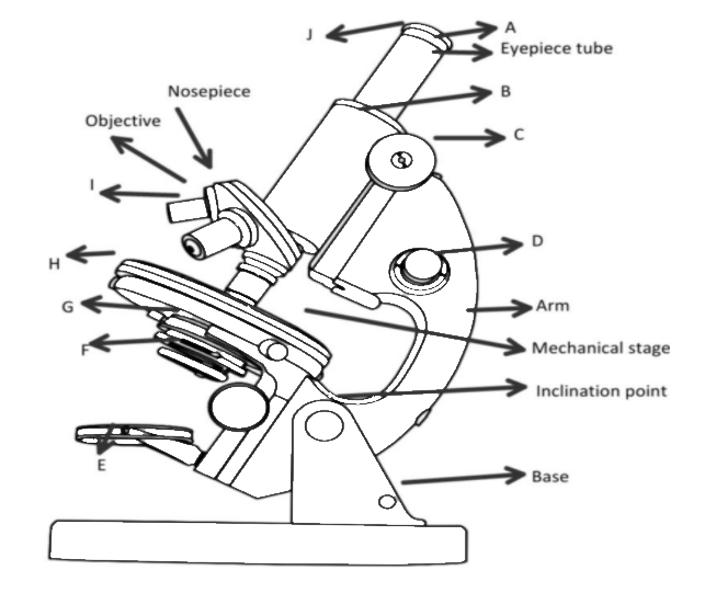
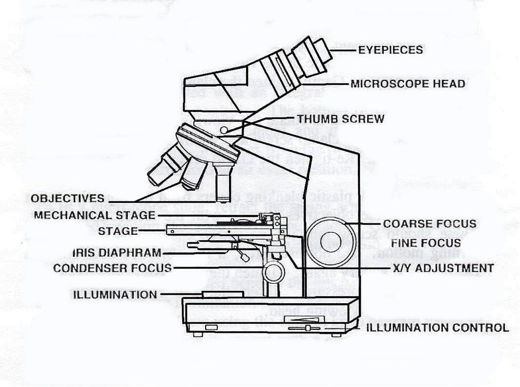
Label Microscope Diagram - EnchantedLearning.com Using the terms listed below, label the microscope diagram. arm - this attaches the eyepiece and body tube to the base. base - this supports the microscope. body tube - the tube that supports the eyepiece. coarse focus adjustment - a knob that makes large adjustments to the focus. diaphragm - an adjustable opening under the stage, allowing ...
Parts of the Microscope Label and Definition Diagram | Quizlet Start studying Parts of the Microscope Label and Definition. Learn vocabulary, terms, and more with flashcards, games, and other study tools. ... Supports microscope. Students also viewed. Similarities and Differences Among Living Organisms. 31 terms. cjenne. Forensic Vocabulary. 11 terms. kschnake1. EntreCulturas 1 Unidad 3.
Microscope, Microscope Parts, Labeled Diagram, and Functions Microscope, Microscope Parts, Labeled Diagram, and Functions What is Microscope? A microscope is a laboratory instrument used to examine objects that are too small to be seen by the naked eye. It is derived from Ancient Greek words and composed of mikrós, "small" and skopeîn,"to look" or "see".
Types of Microscopes: Definition, Working Principle, Diagram ... - BYJUS Stereo Microscope Diagram Principle of Stereo Microscope A stereo microscope works on the reflected light from the sample. The magnification of the microscope takes place at low power and hence, it is suitable for magnifying opaque objects. It is suitable for thick and solid samples because it uses light reflected from the sample.
microbenotes.com › parts-of-a-microscopeParts of a microscope with functions and labeled diagram Parts of a microscope with functions and labeled diagram September 17, 2022 by Faith Mokobi Having been constructed in the 16th Century, Microscopes have revolutionalized science with their ability to magnify small objects such as microbial cells, producing images with definitive structures that are identifiable and characterizable.
Microscope Labeled Diagram in 2022 | Basketball skills, Labels, Diagram Marami pa tulad nito. A life science stock art gallery of general biology, human and veterinary anatomy, zoology, botany and wildlife illustration. Do you want to learn how to draw a microscope easy and quick? If so, be sure to open this very simple drawing tutorial and learn how to draw a microscope.
Microscope Parts, Function, & Labeled Diagram - slidingmotion Microscope Parts Labeled Diagram The principle of the Microscope gives you an exact reason to use it. It works on the 3 principles. Magnification Resolving Power Numerical Aperture. Parts of Microscope Head Base Arm Eyepiece Lens Eyepiece Tube Objective Lenses Nose Piece Adjustment Knobs Stage Aperture Microscopic Illuminator Condenser Lens
Parts of a microscope with functions and labeled diagram Web17. Sept. 2022 · Figure: Diagram of parts of a microscope. There are three structural parts of the microscope i.e. head, base, and arm. Head – This is also known as the body. It carries the optical parts in the upper part of the microscope. Base – It acts as microscopes support. It also carries microscopic illuminators.
16 Parts of a Compound Microscope: Diagrams and Video The 16 core parts of a compound microscope are: Head (Body) Arm Base Eyepiece Eyepiece tube Objective lenses Revolving Nosepiece (Turret) Rack stop Coarse adjustment knobs Fine adjustment knobs Stage Stage clips Aperture Illuminator Condenser Diaphragm Video: Parts of a compound Microscope with Diagram Explained
Label Microscope Diagram - EnchantedLearning.com Label Microscope Diagram Using the terms listed below, label the microscope diagram. Inventions and Inventors arm - this attaches the eyepiece and body tube to the base. base - this supports the microscope. body tube - the tube that supports the eyepiece. coarse focus adjustment - a knob that makes large adjustments to the focus.
Governor Newsom Signs Sweeping Climate Measures, Ushering in … Web16. Sept. 2022 · New California laws will create 4 million jobs, reduce the state’s oil use by 91%, cut air pollution by 60%, protect communities from oil drilling, and accelerate the state’s transition to clean…
Cardiac Muscle Under Microscope with Labeled Diagram Cardiac Muscle Under Microscope with Labeled Diagram 17/12/2022 17/12/2022 by anatomylearner The cardiac muscle under a microscope shows a short cylindrical fiber with a centrally placed oval nucleus. You will find some unique features in cardiac muscle that will help you to differentiate it from skeletal and smooth muscles.
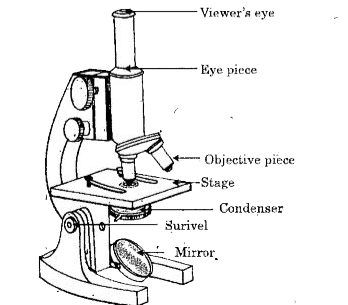
Labelled Diagram of Compound Microscope The below mentioned article provides a labelled diagram of compound microscope. Part # 1. The Stand: The stand is made up of a heavy foot which carries a curved inclinable limb or arm bearing the body tube. The foot is generally horse shoe-shaped structure (Fig. 2) which rests on table top or any other surface on which the microscope in kept.
PDF Label parts of the Microscope Label parts of the Microscope: . Created Date: 20150715115425Z
Microscope Labeling - The Biology Corner The labeling worksheet could be used as a quiz or as part of direct instruction where students label the microscope as you go over what each part is used for. The google slides shown below have the same microscope image with the labels for students to copy.
Diagram of a Compound Microscope - Biology Discussion 1. It is noted first that which objective lens is in use on the microscope. 2. Stage micrometer is positioned in such a way that it is in the field of view. 3. The eyepiece is rotated so that the two scales, the eyepiece or ocular scale and the stage micrometer scale, are parallel. 4.
Microscope Labeling Diagram | Quizlet Hold the slide in place on the stage. Nosepiece Holds the objective lenses and allows the lenses to rotate for viewing. Stage Supports the slide where the specimen is being viewed. Lamp Projects or reflects light upward through the diaphragm. Base Supports and stabilizes the microscope. Diaphragm
Microscope: Parts Of A Microscope With Functions And Labeled Diagram. Figure: A diagram of a microscope's components The microscope has three basic components: the head, the base, and the arm. Head:Occasionally, the head is considered the body. It holds the optical components of the upper part of the microscope. Base:The microscope's base provides great support. It is also equipped with miniature illuminators.













Post a Comment for "44 microscope label diagram"